Volume 2
-
No. 12 December 2000
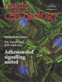
cover image Fluorescence photograph of paxillin (red) and actin (green) in kidney epithelial cells. Nuclei are shown in blue. [Cover design: Majo Xeridat] [review, pE231]
-
No. 11 November 2000
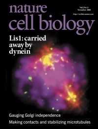
Association of the lissencephaly gene product LIS-1 with mitotic kinetochores. Immunofluorescence image of a mitotic MDCK cell, showing microtubules in red, DNA in pink and LIS-1 in green.784pE201
-
No. 10 October 2000
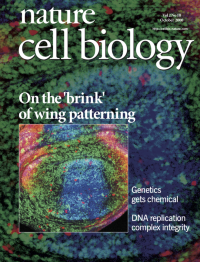
Drosophila wing imaginal disc. Several clones of cells lacking the schnurri product are shown by the absence of green fluorescent protein expression. In these mutant cells, expression of brinker is strongly upregulated, whereas spalt expression (as indicated by blue colouration) is lost. [Cover design: Majo Xeridat] [article, p745]
-
No. 9 September 2000
Extensive branching of cellular processes, induced by overexpression of p120 catenin (green) in NIH3T3 cells. Actin filaments are shown in red. [Cover design: Majo Xeridat] [article, p.637]
-
No. 8 August 2000
Deconvolution image of endogenous mPAR-6 in dissociated murine cortical neurons isolated from 16.5-day-old embryos. Eight hours after dissociation and plating, cortical neurons were fixed and stained with a polyclonal antibody raised against mPAR-6 (red). Neurons were counterstained with Heochst 333298 (blue). Endogenous mPAR-6 is enriched in perinuclear structures and filamentous structures in developing growth cones.
-
No. 7 July 2000
cover image Video confocal analysis of a human osteocarcinoma cell stably expressing GFP–actin 12 h after infection with vaccinia virus. Consecutive video frames 10 s apart are false-coloured blue, green and red to facilitate visualization of the actin-based movement of the virus. The video sequence was recorded during the 1999 EMBO course on ‘Biophysical and mathematical approaches to cell biology’ at EMBL by E. Stelzer, F. Frischknecht and M. Way. [Cover design: Majo Xeridat] [article, p.441]
-
No. 6 June 2000
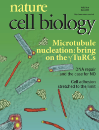
Montage of images that together provide evidence that the γ-tubulin ring complex (γTURC) forms a helical template for microtubule nucleation. These include: immunofluorescence images of microtubules (red) with ends labelled for γTURC components (green); electron micrographs of microtubules (cyan), minus-end labelled with gold particles (yellow) conjugated to antibodies against γTURC components (antibody 'shell' shown as red rings); and tomographic reconstruction of a purified Drosophila γTURC (light green). [Cover design: Majo Xeridat] [article, p. 352, 358 and 365; news and views, p.E93].
-
No. 5 May 2000
3D reconstruction of a polytene nucleus in an east overexpressing larval salivary gland. Spread chromosomes are pale orange, and EAST protein accumulating in intervening extrachromosomal spaces is darker orange. [Cover design: Majo Xeridat] [article, p.268; news and views, p.E74]
-
No. 4 April 2000
An enlargement of a stage 10 wild-type Drosophila oocyte: oocyte is to the right, nurse cells to the left, follicle cells on the top and bottom of the oocyte. The Swallow protein (stained in blue) is asymmetrically localized to the anterior pole of the oocyte. [Cover design: Majo Xeridat] [article, p.185; news and views, p.E60]
-
No. 3 March 2000
Human cells (HeLa) undergoing apoptosis after exposure to TNF. These cells express cytochrome fused to green fluorescent protein (GFP). After treatment, cytochrome-GFP (green) moves from mitochondria to the cytosol. The cells round, then bleb and externalize phosphatidylserine, identified by association with annexin V (red). Finally, a DNA dye (blue) stains the nuclei after the collapse of plasma-membrane integrity.
-
No. 2 February 2000
cover image Synchronous chromosome segregation in an early Drosophila embryo. Spindle microtubules are shown in red, DNA is in green, and the centrosomal protein g-tubulin is in blue. DNA damage and DNA-replication defects trigger mitosisspecific dissociation of g-tubulin from the centrosome and block chromosome segregation. [Cover design: Majo Xeridat] [article, p.90; news and views, p.E28]
-
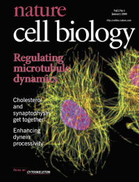
No. 1 January 2000
XMAP215 is one of the major microtubule-stabilizing proteins in Xenopus, and plays an important part in regulating microtubule dynamics. The cover shows interphasic Xenopus XL177 cells, in which XMAP215 (red) binds to microtubules (green; overlay with XMAP215 is yellow). Blue identifies DNA. [Cover design: Majo Xeridat][article, p.13; news and views, p. E11]
Focus