Volume 1
-
No. 8 December 1999
A confocal section of a Drosophila wing imaginal disc. The anterior compartment (left) is wild-type; the posterior compartment (right) is transgenic (Dakt1 has been ectopically expressed with engrailed–GAL4). This confocal section is stained with Hoechst dye for nuclei (blue), rhodamine-conjugated phalloidin for actin (red) and FITC for Dakt1 (green). Note the larger size of cells ectopically expressing Dakt1. [Cover design: Majo Xeridat] [article, p. 500; news and views, p.E191]
-
No. 7 November 1999
In this picture of live budding yeast cells, the fluorescence of green fluorescent protein (GFP) fused to the DNA-replication factor Mcm4 has been merged with a phase-contrast image. Mcm4–GFP accumulates in the nucleus at the end of mitosis, and is subsequently excluded from the nucleus by cyclindependent kinase activity, as bud emergence and DNA replication occur in the next cell cycle. [Cover design: Majo Xeridat] [article, p. 415; news and views, p.E175]
-
No. 6 October 1999
Dendritic cells in the process of delivering internalized antigen to the cytosol. Cells internalized antigen for 1 h. The middle and lower cells were then incubated for 3 or 6 h more, respectively, at 37°C. Staining was with anti-rabbit IgG (green) for internalized antigen and lysotracker (red) for acidic endocytic compartments.Yellow indicates presence of antigen in acidic vesicles. [Cover design: Majo Xeridat] [article, p.362.; news and views, p. E152.]
-
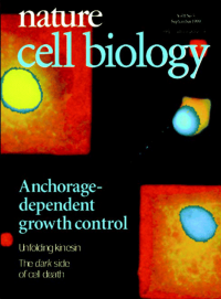
No. 5 September 1999
cover imagePseudocoloured fluorescence micrograph showing local shapedependent growth control in endothelial cells grown on fibronectin-coated adhesive islands of different sizes (red). Highly spread cells enter S phase (yellow); smaller, retracted cells remain quiescent (blue) in the same medium. Image: courtesy of Christopher Chen and Don Ingber.)[Cover design: Majo Xeridat]p. E131
-
No. 3 July 1999
Confocal section through the epithelium of a Drosophila embryo at the blastoderm stage. Staining with an anti-E-APC antiserum (blue) and with an anti-Armadillo antiserum (pink) merge at zones enriched in adherens junctions (yellow). [Cover design: Majo Xeridat] [articles, p. 144]
-
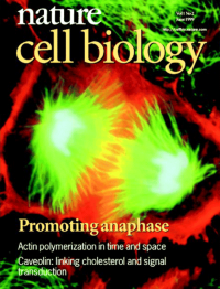
No. 2 June 1999
cover imagePtK1 cell in late anaphase soon after the onset of cytokinesis. The cell was fixed, then stained with phalloidin to label the filamentous actin (red), DAPI to label the DNA (pale blue), and anti-alpha-tubulin antibodies to label the microtubules (green). (Image: courtesy of Julie Canman and Ted Salmon.) [review, p.E47]
-
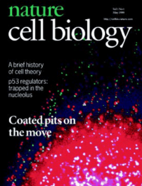
No. 1 May 1999
In the periphery of an intact cell, GFPlabeled clathrin coated pits (red) are retained and shift coordinately upon detergent extraction (green) suggesting attachment to an interconnected, underlying membrane skeleton. [articles, p. 1]