
Volume 17 Issue 11, November 2012
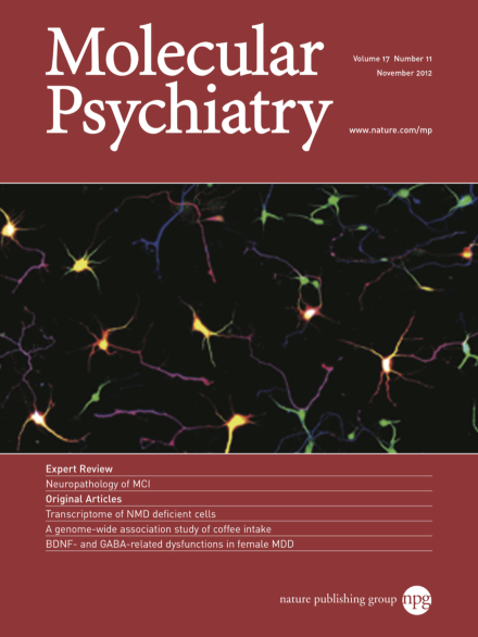
Image of control mouse primary hippocampal neurons used to compare the effect of ARHGAP24 over-expression. Isolated neurons were transfected with EGFP (psudeocolouredred) and grown for 4 days in-vitro before fixation. Immuno-fluorescent staining using anti-MAP2 (psuedo-green) and anti-TAU1 (psuedo-blue) antibodies identified dentrites and axons respectively. For more info on this topic, please refer to the article by Nguyen et al. on pages 1103–1115.
Image
-
Advertisement
