
Volume 12 Issue 2, March 2019
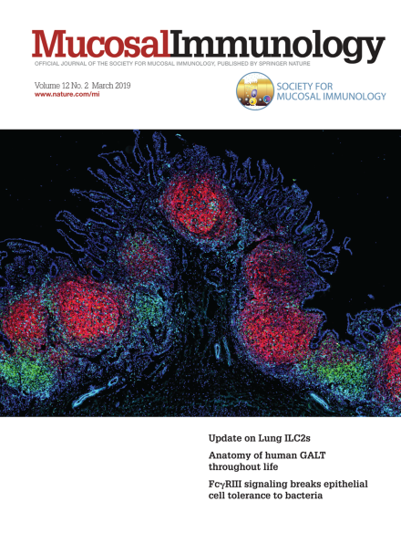
This image shows a high density of lymphoid structures in Peyer’s patches of the human ileum from an 18 month old pediatric organ donor. Frozen intestinal tissues were stained for expression of CD4 (green), CD8 (blue), and CD45RA (red) for distinguishing T cells and B cell follicles, and DAPI (cyan) to visualize nucleated cells.
For further information see article in this issue, page 380.Image Courtesy of Takashi Senda et al., Columbia Center for Translational Immunology, Columbia University in the City of New York.
Editorial
-
Advertisement
