
Figures, tables and video
From the following article
Jyoti N. Sengupta
GI Motility online (2006)
doi:10.1038/gimo16
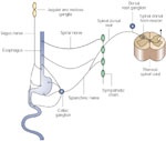
Figure 1
Schematic diagram of vagal and spinal nerve supply to the esophagus.
Full size figure and legend (44K)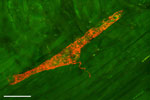
Figure 2
Confocal photomicrograph of anterogradely labeled intraganglionic laminar ending (IGLE) in the proximal striated part of the esophagus of the rat.
Full size figure and legend (80K)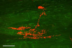
Figure 3
Confocal photomicrograph of a vagal intramuscular arrays (IMA) nerve terminal in a tangential section of the lower esophageal sphincter of a rat.
Full size figure and legend (71K)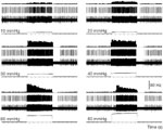
Figure 4
Intensity-dependent response of a vagal afferent fiber to graded esophageal distention (ED, 5–80 mmHg, 30 seconds).
Full size figure and legend (85K)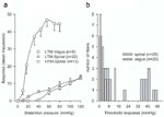
Figure 5
Comparison of mechanosensitive properties of vagal and spinal afferent fibers innervating the esophagus of opossum (Didelphis virginiana).
Full size figure and legend (35K)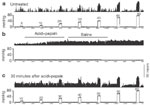
Figure 6
Response of a vagal distention-sensitive afferent fiber to graded esophageal distention (ED) before and after acid infusion.
Full size figure and legend (58K)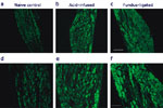
Figure 7
Transient receptor potential vanilloid-1 (TRPV1)–like immunoreactivity (TRPV1–IR) in rat nodose ganglia.
Full size figure and legend (39K)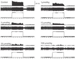
Figure 8
Effect of N-methyl-D-aspartate (NMDA) open channel blocker memantine hydrochloride on mechanotransduction property of an esophageal vagal afferent fiber innervating the esophagus of rat.
Full size figure and legend (66K)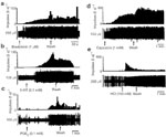
Figure 9
Response characteristics of mucosal mechanosensitive afferent fibers.
Full size figure and legend (72K)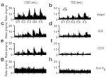
Figure 10
Responses of a distention-responsive upper cervical (C2) spinal neuron having convergent inputs from the cervical and thoracic esophagus.
Full size figure and legend (49K)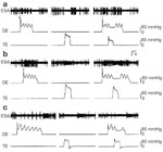
Figure 11
Typical firing patterns of three types of neurons in the nucleus tractus solitarius centralis (NTSc) region receiving synaptic input from the vagal afferent fibers.
Full size figure and legend (57K)