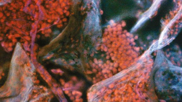
Thank you for visiting nature.com. You are using a browser version with limited support for CSS. To obtain
the best experience, we recommend you use a more up to date browser (or turn off compatibility mode in
Internet Explorer). In the meantime, to ensure continued support, we are displaying the site without styles
and JavaScript.
Advances in microscopy and the application of machine learning to histology will modernize the examination of tissues in the clinical laboratory and in the operating room.
Graph deep learning can leverage information in the tumour microenvironment to extract prognostic histopathological features from gigapixel-sized whole-slide images.
A data-efficient and interpretable deep-learning method for the multi-class classification of whole-slide images that relies only on slide-level labels is applied to the detection of lymph node metastasis and to cancer subtyping.
Deep learning can be used to virtually stain autofluorescence images of unlabelled tissue sections, generating images that are equivalent to the histologically stained versions.
A slide-free, inexpensive and non-destructive microscopy technique rapidly provides high-resolution histology images that resemble those obtained from conventional haematoxylin-and-eosin-stained specimens.
A method that identifies patterns of tumour heterogeneity in intact biopsy samples using 3D light-sheet microscopy stratifies patients by tumour stage.
A light-sheet microscope images large surgical and biopsy specimens non-destructively over large fields of view in two and three dimensions, with the same level of detail as traditional slide-based histopathology.
By taking advantage of stimulated Raman spectroscopy and fibre-laser technology, virtual histology images can be obtained in real time in the operating room, with diagnostic quality comparable with that achieved via conventional histopathology.
Weakly supervised deep-learning models for the analysis of whole-slide images from tumour biopsies perform better at prognostic tasks if the models incorporate context from the local microenvironment.
A deep-learning model for cancer detection trained on a large number of scanned pathology slides and associated diagnosis labels enables model development without the need for pixel-level annotations.
Optical imaging of fluorescently labelled tissue illuminated by ultraviolet light does not require microscope slides and makes for a rapid alternative to conventional histology.
Stimulated Raman spectroscopy combined with machine learning generates histological images for the rapid diagnosis and classification of brain tumours.