
Visiting the dental practice is a valuable opportunity for skin cancer screening, says Ben J. Steel.

Skin cancer is the commonest form of cancer in the UK. In 2010, 112,367 skin cancers were registered in the UK out of a total of 424,128 cancers of all types.1 Three main types are recognised: basal cell carcinoma (BCC) and squamous cell carcinoma (SCC), which collectively comprise non-melanoma skin cancer (NMSC), and malignant melanoma (MM).

Examination of the oral mucosa for signs of oral squamous cell carcinoma and other mucosal conditions is an accepted part of the normal dental check-up, as is an extra-oral examination to check the facial hard and soft tissues, jaw joints and cervicofacial lymph nodes.2 A brief look at the head and neck skin for suspicious lesions could easily be incorporated into this structure. In the two years leading to September 2012, 29.6 million people in the UK attended a dentist, representing some 52.1% of the adult population.3 With such a considerable proportion of the population passing through, there exists a valuable opportunity for a form of ad hoc screening for head and neck skin malignancy. Patients with suspicious lesions could be referred to their general medical practitioner (GP) or an oral and maxillofacial surgeon for further management.
This article will present an overview of the three forms of skin cancer most likely to be seen among dental patients in the UK.
Basal cell carcinoma
Epidemiology
This is the commonest type of skin cancer, and indeed any cancer, in the UK, with at least 48,000 cases registered in England each year between 2004 and 2006.4 This is believed to be an underestimate, with the real number of cases likely to be 55,000-60,000 a year.5 It is becoming more common – increasing by 1.4% and 1.9% for males and females respectively between 1992 and 2003, which was most marked in the 30-39 age group.6 It is known that having one BCC increases the likelihood of having other lesions, both at the same time and later.7 There is regional variation – the age-standardised incidence in the south west of England is 121.3 per 100,000 compared to 93.7 across England as a whole.8 The incidence increases with age, above the age of 55 having a male predominance.
Pathology and risk factors
BCC is a malignant neoplasm of keratinocyte stem cells. It behaves as a slowly growing locally destructive neoplasm, which may over time infiltrate underlying tissues such as bone and cartilage; however, metastasis is extremely rare. If neglected BCCs may reach a substantial size and deaths have been reported, albeit very rarely. The vast majority of cases are sporadic and caused by ultraviolet (UV) radiation, specifically UV-B.9. Both chronic and acute exposure (as sunburn) are implicated, which damage DNA via both direct effects and indirectly via production of oxygen or nitrogen radical species, or induction of inflammation. As well as age and male gender, having fair skin is a risk factor, as is having fair or red hair, or freckles. People of Celtic ancestry are most at risk. Most cases occur on sun-exposed areas such as the face and neck, scalp, forearms, hands, lower legs and feet.8 Intraoral lesions are vanishingly rare, and when reported often thought to have been misdiagnosed.10
Several genetic conditions predispose to BCCs, notably nevoid basal cell carcinoma syndrome (NBCCS), also known as Gorlin-Goltz syndrome. Typical manifestations are multiple, aggressive BCCs occurring from a young age, keratinising odontogenic tumours, skeletal abnormalities and plantar and palmar pits. It is thought around 2% of people with BCCs before the age of 45 have this condition.11 Other rare conditions include Bazex-Dupré-Christol syndrome and Rombo syndrome.
Clinical presentation
BCCs typically cause no symptoms and may not be noticed by the patient. Three main variants are recognised: noduloulcerative, superficial and infiltrating.
The noduloulcerative type represents the typical and most common form of BCC. The appearance is initially a firm red or pink painless papule, which slowly enlarges and forms a central area of ulceration surrounded by a raised semi-translucent margin with telangiectasia (visible tiny blood vessels coursing through it) (Fig. 1). At this stage it is also known as a rodent ulcer. The lesion may bleed occasionally but does not heal. A pigmented variant shares the same morphology but is pigmented by melanin deposition – appearing tan, brown, black or bluish. The pigment is usually not distributed evenly, whereas that in a benign melanocytic lesion (mole) would be.

Noduloulcerative BCC
Superficial type lesions are the second most common form, and occur more often on the trunk, appearing as flat or raised, red or pink, scaly eczematous areas, sometimes with ulceration or crusting (Fig. 2). It may be darkly pigmented. The borders are well-demarcated and can be slightly raised or rolled, with a ’threadlike’ appearance. They may be mistaken for a patch of eczema or psoriasis and can be multiple.

Superficial BCC
Infiltrating lesions are insidious lesions and less common than the other forms, tending to be more aggressive. Their appearance mimics scar tissue, being flat skin-coloured, pink or whitish lesions that are firm to palpation. The borders are indistinct and may be slightly elevated (Fig. 3). Their insidious nature means there may be more invasion before they are noted than the other forms.

Infiltrating BCC
Investigations and treatment
BCCs with a typical appearance are generally not biopsied and are instead diagnosed clinically. Treatment depends on the location, size and histology of the lesion. Surgical excision is most commonly employed, either with direct edge apposition or local flap closure, typically under local anaesthetic. This may be done by GPs in primary care, or by a range of specialties in hospital. A 4-5 mm surgical margin gives a 95% cure rate,12 although infiltrating and large BCCs are thought to require larger margins. For larger lesions, or where surgery is declined or inappropriate, radiotherapy alone can be employed. Mohs micrographic surgery, a technique where tissue samples are viewed microscopically during the operation, can be used to ensure complete excision of recurrent or poorly-defined lesions. A range of other modalities – curettage, electrocautery, cryotherapy, photodynamic therapy or carbon dioxide laser ablation, are sometimes used to destroy low-risk lesions, but are not recommended for high-risk lesions.13 Imiquimod is an immune response modifier, used topically as a 5% cream applied to the lesion five times a week for six weeks, and appears to be effective in treating small superficial lesions.14
A new treatment licensed in the UK in August 2013 is the biological agent Vismodegib. It is only used in locally advanced tumours whose position prevents surgical removal, or if recurrence has occurred after resection and radiotherapy has already been used. Its use is not yet widespread but early results are promising.15
Differential diagnosis
-
Actinic keratosis – see SCC section
-
Seborrhoiec keratosis – common, usually asymptomatic, benign neoplasm, of variable appearance but usually beginning with a sharply defined light brown macule, later growing a velvety, verrucous, or warty surface with keratin plugs and coloured pale brown to black (Fig. 4). Slowly grows over time, does not resolve and does not malignantly transform, although the presence of multiple lesions may make a malignancy difficult to notice. Can also be confused with SCC and MM. No treatment is necessary
Figure 4 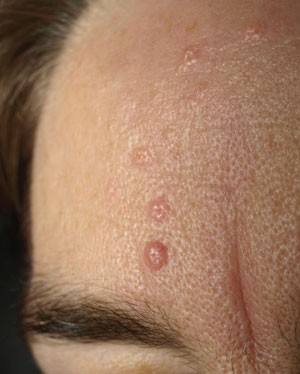
Seborrhoeic keratosis
-
Angiofibroma
-
Bowen disease – see SCC section
-
Fibrous papule of the face
-
Molluscum contagiosum
-
Psoriasis
-
Melanocytic naevus
-
SCC, MM or metastatic malignancy
-
Cutaneous T cell lymphoma
-
Darier disease
-
Eczema
-
Keratoacanthoma – see SCC section
-
Lichenoid keratosis.

Squamous cell carcinoma
Epidemiology
This is the second most common skin cancer in the UK and worldwide. Reported age-standardised incidences for cutaneous SCC range from around 16 per 100,000 in London to around 36 per 100,000 in the south west of England, equating to at least 10,000 cases per year in England and Wales (compared to around 5,000 cases of intraoral SSCs).1 Data suggest an increase in incidence in recent years.13
Pathology and risk factors
SCC is a malignant neoplasm of keratinocytes that occurs predominantly in sun-exposed areas of the body, but can occur on any skin or mucosal surface. It is prone to invade locally and metastasise. SCC pathogenesis is a stepwise process, in that a number of sequential mutations of proto-oncogenes and tumour suppressor genes are needed for carcinoma to be induced. The main risk factor for its development is UV radiation. Chronic exposure, including from sunbeds,16 is mainly implicated,17 with acute exposure thought to be less important.18 Human papillomavirus is a suspected risk factor19 although the evidence is not as strong as for other sites such as the cervix and oropharynx. Increased age, male gender and Caucasian ethnicity are associated with increased risk, as are irradiation and exposure to chemicals such as arsenic and polycyclic aromatic hydrocarbons. A recent meta-analysis19 found no association with cigarette smoking (unlike for oral mucosal SCC). It is known that transplant recipients and the immunosuppressed (through drugs or disease) are at increased risk; its magnitude increases with the degree and chronicity of the immunosuppression. Genetic conditions such as xeroderma pigmentosum (defective DNA repair mechanisms after UV exposure) and oculocutaneous albinism (reduced or absent skin pigmentation) also carry an increased risk of SCCs.
Clinical presentation
SCCs may present as a new enlarging ulcer, lump or red patch on the skin, changes to a pre-existing lesion or as a sore that does not heal. There is typically no pain although there may be mild tenderness to touch. Rarely, with perineural involvement there may be localised pain, numbness, muscle weakness or twitching. In the head and neck this may manifest as cranial nerve deficits.
The appearance of SSCs is very variable but the classic presentation is a raised indurated ulcer, with rolled margins, sometimes with an erythematous periphery (Fig. 5). Other surface changes include scaling, crusting and a cutaneous horn (a very thickened raised area of keratin) (Fig. 6). Less commonly it may be a nodule with an intact pinkish surface. The most common sites are the lower lip, ear pinnae, pre-auricular regions, forehead and scalp. There may be palpable lymph nodes, in this context particularly the pre-auricular/parotid and upper cervical nodes. Various histiological subtypes are recognised, but around 60% of invasive skin SCCs appear within a pre-existing actinic keratosis.20

Ulcerative SCC on the pinna

Cutaneous horn presentation of SCC
Actinic keratosis, also known as solar keratosis, is premalignant for SCC, and is caused by exposure to UV radiation. It is common in adults – 23% of over 60s had at least one lesion in one UK study,21 although is rare under 40 years. It is asymptomatic but may cause some mild irritation or itching. Clinically it is an irregular scaly keratotic plaque, coloured from pink to white or grey, sometimes with an erythematous background, and is often multiple (Fig. 7). The commonest sites are the face, scalp of bald men, forearm and dorsum of the hand. It is often easier to feel the sandpaper-like surface of these lesions than to see them. Histologically it encompasses a range from cellular atypia at the basal layer only, to carcinoma in situ, where there is full thickness atypia and abnormal maturation. The risk of progression to invasive SCC has been variously quoted at between 0.25 and 20% a year.22 The same process occurring on the lip is known as actinic cheilitis. Changes indicative of malignant transformation within an actinic keratosis include development of areas with sharp circumscription, increase in thickness or the degree of scaling, and formation of an exophytic lesion, indurated (hardness) or, particularly, the development of ulceration.

Actinic keratosis on the scalp
Investigations and treatment
The diagnosis of SCC is confirmed by biopsy, and the tumour graded and staged based on the size and involvement of underlying structures and lymph nodes, and the presence or absence of distant metastases. Clinically positive lymph nodes are biopsied by fine needle aspiration or excisional node biopsy, and if neoplastic cells are present, regional lymph node dissection undertaken. Assessment for metastasis may be undertaken with computer tomography (CT) or positron emission tomography (PET) scans. SCC occurring within actinic keratosis is generally low risk, with a low risk of metastasis and a favourable prognosis.20
Standard treatment is surgical excision, with the margin determined by the risk status of the tumour. Low risk, smaller (less than 2 cm diameter) tumours are excised with a 4 mm margin, and higher risk (tumours wider than 2 cm or at high risk site – ear, scalp, eyelids or nose) with a 6 mm margin.23 Mohs micrographic surgery, as previously described, leads to less recurrence than following excision alone.24 Curettage or cautery may be used for small well-differentiated tumours, but guidelines advise cryotherapy be used with caution.25
Radiotherapy may be used as a sole treatment for smaller lesions, particularly in areas of cosmetic and functional sensitivity, for example, lower eyelid, medial canthus, lip, although is contraindicated in younger patients as surgical scars usually have a better cosmetic outcome. Post-operative radiotherapy is also used as an adjuvant when the excised specimen shows close margins, perineural invasion or other high-risk histological features.
Chemotherapy is used in selected cases following surgical resection or in unresectable disease, typically with regimens including cisplatin or 5-fluorouracil. Cetuximab is a well-tolerated newer agent licensed in the UK for use in locally invasive SCC of the head and neck in combination with radiotherapy.
Actinic keratoses do not always require treatment – decisions are made based on individual circumstances. Cryotherapy, photodynamic therapy, or topical immunomodulator, diclofenac or keratolytic creams are frequently used modalities.26
Differential diagnoses
-
Actinic keratosis – see above
-
Keratoacanthoma – this has variously been considered as a distinct entity or a well-differentiated form of SCC. It presents on sun exposed skin, typically in elderly persons with fair skin, as a rapidly enlarging crateriform nodule that may reach 2 cm or so in diameter, becoming dome shaped with central ulceration and typically lacking induration or fixation to underlying tissue (Fig. 8). Over subsequent months it generally spontaneously involutes to leave a scar, although aggressive spread and metastasis has occasionally been observed. Microscopically it can be difficult to distinguish from SCC, and definite histological criteria for keratoacanthomas and keratoacanthoma-like SCCs are lacking.27 Due to diagnostic difficulties these are often excised in the same way as other SCCs
Figure 8 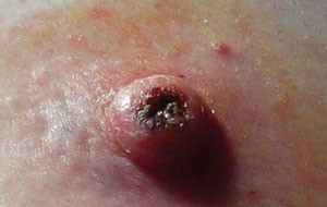
Keratoacanthoma
-
Bowens disease – an uncommon in situ SCC, presenting as an irregular well-demarcated erythematous scaly keratotic plaque on sun-exposed skin, which is generally asymptomatic
-
Seborrhoiec keratosis
-
Atopic dermatitis
-
Atypical fibroxanthoma
-
Congenital skin tumours, for example, dermoid/dermolipoma
-
Chemical burns
-
Contact dermatitis
-
Pyoderma gangrenosum
-
BCC.

Malignant melanoma
Epidemiology
MM is the fifth most common cancer in the UK, with 12,818 cases registered in the UK in 2010, and a UK-wide age-standardised incidence of 17.1 per 100,000.27 The UK distribution is fairly even although it occurs more in the south west of England. The age distribution is younger than for other skin cancers. Incidence increases steadily with age, with a female predominance below age 55 (and a female:male ratio in the age group 20-24 of 10:4) and male predominance above 55. The number of MM cases in the UK increased by 55% between 2000 and 2010. Twenty-two percent of lesions in males occur in the head and neck area, compared to 14% in females.27
Pathology and risk factors
MM is a malignant neoplasm of melanocytes that can arise within a pre-existing benign melanotic lesion or de novo from apparently normal skin. It tends to invade locally and metastasise, and can occur at any skin site, or the oral, genital, ocular or urinary epithelial surfaces.
UV radiation, especially UV-B, is the main risk factor. Recent studies suggest MM of the head and neck is more strongly related to sunburn, and of the trunk to more chronic sun exposure.28,29 People with fair skin, blue eyes, fair or red hair and those who burn easily are at increased risk of MM, as are those with freckling and benign naevi, especially those with more than 100 such lesions. Large or giant congenital melanocytic lesions confer a high risk, as does the presence of any atypical or dysplastic naevi, or if MM itself has previously occurred. Immunosuppression - therapeutic for solid organ transplant or pathological in lymphoma and HIV/AIDS - increases the risk, although the effect of antiretroviral treatment is unclear.30 Family history is also important – those with a first-degree relative with MM are at increased risk,31 and specific inherited conditions confer an additional risk so as to justify routine screening by a dermatologist from childhood – dysplastic naevus syndrome and xeroderma pigmentosum. Increased overseas holidays, sunbathing, use of sunbeds32 and outdoor recreational behaviour is thought to be responsible for most of the rise in cases in recent years.28
Clinical presentation
MM typically does not cause any symptoms and may be noticed as a new lesion on the skin or as a change in a pre-existing lesion. Three main morphological and many histological subtypes of MM are seen in the head and neck. The relationship between these morphological and histological variants is not always clear.21
The superficial spreading form comprises 70% of skin MMs,21 and presents as a macular (flat) or slightly raised lesion that can be a variety of colours (brownish, grey, black, blue, whitish or pink). It is typically less than 3 cm in diameter although can grow much larger than this. The presence of induration or surface nodules within these lesions indicates likely deeper invasion (Fig. 9), as does the formation of satellite pigmented areas around the original lesion.

Superficial spreading MM
The nodular form comprises around 15% of MMs and is an exophytic nodular lesion, usually deeply pigmented and often asymmetrical, that may be ulcerated (Fig. 10). Occasionally these are so poorly differentiated that pigment is absent, instead appearing pinkish red – an amelanotic melanoma. It tends to grow in a more vertical pattern, in contrast to the horizontal pattern shown by the superficial spreading form.

Nodular MM
Lentigo maligna melanoma comprises 5-10% of MMs and develops within a lentigo maligna, which is an in situ melanoma occurring on sun-exposed skin. It is most common on sun-exposed skin in the elderly. Clinically, lentigo maligna resembles a superficial spreading melanoma, being a large, flat lesion with irregular borders, and shares the same variety of colours (Fig. 11). It enlarges slowly over many years, with the development of nodularity or induration indicating a change to invasive melanoma. The risk of progression from lentigo maligna to MM is controversial but thought to be 4.7% or less over a lifetime.33

Lentigo maligna
A useful rule to judge the likelihood of malignancy in pigmented lesions is the ABCDE rule (Table 1). The presence of one or more of these features may raise suspicion of a malignancy.
Investigations and treatment
Any lesion where MM is suspected is photographed and subjected to excisional biopsy with a 2 mm margin, in order to confirm the diagnosis of MM and provide information about the tumour thickness (termed the Breslow thickness), and other histological parameters of prognostic importance. Incisional or punch biopsies are generally not used. Following definitive diagnosis, wide local excision of the area around the biopsy site is required – the Breslow thickness is used to determine the surgical margin required. With a thickness of 1 mm, a 1 cm margin is used, for 1-2 mm a 1-2 cm, 2-4 mm a 2-3 cm and more than 4 mm a 3 cm margin.34
Sentinel node biopsy (SNB) may be considered where Breslow thickness exceeds 1 mm and no nodes are clinically evident. This involves injection of a radiolabelled substance, and later a blue dye, close to the tumour, and then identification and biopsy of the node(s) showing the greatest uptake. Around 20% of SNBs are positive, and many of these patients may go on to have completion lymphadenectomy. The role of SNB is controversial, as no survival benefit has been demonstrated,35 and it is not universally employed in the UK. There is no benefit to elective neck dissection in MM.
Clinically-evident nodes are investigated with fine needle aspiration biopsy and treated, if positive, with formal block dissection. When there is no evidence of nodal disease (stages I and II) no further imaging investigations are required, otherwise whole body CT, and sometimes a PET scan, may be used to search for metastases, which, if present, may be treated with surgical excision, or carbon dioxide laser ablation if on the surface. There is some evidence that adjuvant radiotherapy improves local control, although not survival. Conventional chemotherapy generally gives no survival benefit, although a small effect on overall and relapse-free survival has been noted for interferon.
Research into signal transduction pathways involved in MM pathogenesis is leading to new biological therapies targeted to specific identified genetic mutations.36
Prognosis
The 5-year-survival for patients with melanomas of Breslow thickness less than 1 mm is 97% (stage IA), falling to 81% with thickness up to 4 mm (stage IIA), 40% with palpable lymph node involvement (stage IIIC), and around 20% with widespread disseminated disease (stage IV).21
Differential diagnoses
-
Benign melanocytic naevus – commonly known as a ‘mole’, either congenital (non-neoplastic malformation) or acquired (benign neoplasm), almost universal among the white population with incidence peaking between age 40-50. There is an increased risk of malignant transformation to MM, especially with the congenital form, although the incidence is not known. They are macular or slightly raised with uniform colour, and lacking any suspicious features of the ABCDE rule (see Table 2)
Table 2 Suggested approach to skin cancer for dental practitioners -
Dysplastic/atypical naevus – an acquired naevus that is thinly papular and relatively broad, often with a fried egg appearance (more darkly pigmented centre). The term was coined to reflect a perceived high risk of malignant transformation, however, this is controversial, although it is generally believed the risk is higher than a melanocytic naevus – they may represent premalignant lesions or risk factors for de novo lesions or both. Histologically, this lesion is diagnosed if architectural and melanocytic atypia are demonstrated. Dysplastic naevus syndrome is an inherited autosomal dominant condition giving high numbers of benign and dysplastic naevi, with a lifetime risk of MM approaching 100%
-
Lentigo maligna – see above
-
Seborrhoiec keratosis
-
Pigmented actinic keratosis
-
Histiocytoid haemangioma
-
Epithelioid tumour
-
Atypical fibroxanthoma
-
SCC
-
Metastatic tumour.
Clinical approach for the dental practitioner
The suggested role of the dental practitioner is as follows and summarised in Table 2:
-
1
History and risk factor evaluation
-
2
Examination – general and focused
-
3
Counselling on risk factors and self-monitoring
-
4
Onward referral.
History and risk factor evaluation
It is unlikely a patient would volunteer concerns of a skin lesion to the GDP but if this occurs salient points to question in the history include the duration and evolution of the lesion and any features such as pain, itchiness, numbness or surface bleeding. Similar questions would be used to ask about any lesion found on examination. As part of the history-taking process risk factors should be evaluated, in particular personal and family history of skin cancer, sun exposure though work, recreational activities and sunbed use, both chronic and sunburn, medical/drug history and patient complexion.
Examination
To identify lesions a thorough and systematic examination technique is essential. This includes all head and neck skin, paying particular attention to the lower eyelid, nose, ear pinnae, lips, and in bald people, the scalp. Malignant lesions may be very subtle so diligence is required. Any suspicious lesions identified are examined more closely. Important details to note on inspection include: location, size, shape, border, colour, symmetry and uniformity, surface keratin/ulceration/bleeding, and blood vessel pattern and morphology. Palpation follows, to distinguish soft from firm or indurated lesions. Palpation of the regional lymph nodes may reveal lymphatic metastasis, in particular including the pre- and post-auricular, occipital and upper cervical nodes. Rarely, locally invasive lesions may affect facial or trigeminal nerve function, or cause proptosis, diplopia or ophthalmoplegia, which would necessitate formal cranial nerve testing, and require specialist evaluation.
Dermoscopy is an examination technique for skin lesions using good lighting and magnification, typically around 10x. Various commercially-produced devices are available and are commonly used by GPs to identify benign lesions that do not require urgent referral and identify premalignant lesions and BCCs.
Counselling on risk factors
The main modifiable risk factors for skin cancer are chronic and acute sun exposure. British Association of Dermatologists advice37 suggests people should stay out of sunlight during the hottest part of the day (midday to 3 pm), avoid using sunbeds and keep babies and children out of strong sun altogether. A hat and UV-impenetrable clothing should be worn in direct sunlight. Suncream should be at least factor 30 to protect against UV-B and have the circle logo and/or four or five UV-A stars to protect against UV-A, and should be applied 30 minutes before exposure.
Furthermore the patient can be educated at this point about what constitutes a suspicious skin lesion. By having this awareness they are better placed to monitor their own skin and approach their GP early should they notice any suspicious changes.
Onward referral
The 2006 NICE guidelines state that the diagnosis and treatment of all suspicious pigmented lesions, lesions that may be MM, SCC or high risk BCCs, or any lesion where the diagnosis is unclear, ‘should be carried out only be specialists (normally dermatologists)’.38 Precancerous lesions such as actinic keratosis may be, and usually are, managed in primary care by a GP or GP with a special interest, unless there are particular individual circumstances, such as a difficult lesion location, to warrant hospital referral.
In hospital, care is organised by the skin cancer network: either local hospital or specialist skin cancer multidisciplinary teams (MDT). GPs can refer directly into this service and all skin cancer cases, regardless of which speciality the patient is referred to, feed into this MDT. At the MDT meetings these patients are discussed and the management decided. Dentists can refer either to the GP or directly to the oral and maxillofacial surgery department in secondary care. This would be done as an urgent referral under the two-week rule. The interest of OMF surgeons in head and neck skin cancer management varies but many units are active in this area. The best route of referral will therefore vary depending on local circumstances.
Conclusions
Skin cancers are common in the UK, and BCC, SCC and MM are the main forms. Most skin cancers will present as a lesion that has features characteristic of that form, and thus if the clinician is aware of these usual presentations and examines for them, they would be well placed to spot them opportunistically. Such ad hoc screening for head and neck skin cancer fits well into the existing convention for the extra-oral examination a dentist would normally perform. Earlier recognition of suspicious lesions, and informing patients about risk factors and self-monitoring, may lead to improved outcomes.
There is one hour of verifiable CPD associated with this article. To take part, go to: www.nature.com/bdjteamcpd
References
Cancer Research UK. UK Cancer incidence 2010. Cancer Research UK, 2012.
Faculty of General Dental Practitioners at the Royal College of Surgeons of England. Clinical examination and record keeping: good practice guidelines. London: FGDP UK, 2009.
The Information Centre for Health and Social Care. NHS dental statistics for England. 2012–2013 First quarterly report. HSCIC, 2012.
South West Public Health Authority Skin Cancer Hub. Online information available at www.swpho.nhs.uk/skincancerhub/ (accessed March 2014).
Brewster D H, Bhatti L A, Inglis J H, Nairn E R, Doherty V R . Recent trends in incidence of nonmelanoma skin cancers in the East of Scotland, 1992-2003. Br J Dermatol 2007; 156: 1295–1300.
Bath-Hextall F, Leonardi-Bee J, Smith C, Meal A, Hubbard R . Trends in incidence of skin basal cell carcinoma. Additional evidence from a UK primary care database study. Int J Cancer 2007; 121: 2105–2108.
Marcil I, Stern R S . Risk of developing a subsequent nonmelanoma skin cancer in patients with a history of nonmelanoma skin cancer. Arch Dermatol 2000; 136: 1524–1530.
National Institute for Health and Care Excellence. Improving outcomes for people with skin tumours including melanoma (update): the management of low-risk basal cell carcinomas in the community. London: NICE, 2010.
Kim M, Park H J, Baek S C, Byun D G, Houh D . Mutations of the p53 and PTCH gene in basal cell carcinomas: UV mutation signature and strand bias. J Dermatol Sci 2002; 29: 1–9.
Neville B W, Damm D D, Allen C M, Bouquot J E . Oral and maxillofacial pathology. 2nd ed. Philadelphia: Saunders, 2002.
Rahbari H, Mehregan A H . Basal cell naevus epithelioma [cancer in children and teenagers]. Cancer 1982; 49: 350–353.
Breuninger H, Dietz K . Prediction of subclinical tumour infiltration in basal cell carcinoma. J Dermatol Surg Oncol 1991; 17: 574–578.
Holme S A, Malinovszky K, Roberts D L . Changing trends in non-melanoma skin cancer in South Wales. Br J Dermatol 2000; 143: 1224–1229.
Telfer N R, Colver G B, Morton C A . Guidelines for the management of basal cell carcinoma. Br J Dermatol 2008; 159: 35–48.
Amin S H, Motamedi K K, Ochsner M C, Song T E, Hybarger C P . Mechanisms and efficacy of vismodegib in the treatment of basal cell carcinoma. Discov Med 2013; 16: 229–232.
Karagas M R, Stannard V A, Mott L A, Slattery M J, Spencer S K, Weinstock M A . Use of tanning devices and risk of basal cell and squamous cell cancers. J Natl Cancer Inst 2002; 94: 224–226.
Schmitt J, Seidler A, Diepgen T L, Bauer A . Occupational ultraviolet light exposure increases the risk for the development of cutaneous squamous cell carcinoma: a systematic review and meta-analysis. Br J Dermatol 2011; 164: 291–307.
Rosso S, Zanetti R, Martinez C et al. The multicentre south European study ‘Helios’. II: different sun exposure patterns in the aetiology of basal cell and squamous cell carcinomas of the skin. Br J Cancer 1996; 73: 1447–1454.
Birch-Johansen F, Norrild B, Olesen A B, Jensen A, Kjaer S K . HPV infection might play a role in the development of nonmelanoma skin cancer in immunocompetent individuals. Ugeskr Laeger 2012; 174: 413–417.
Song F, Qureshi A A, Gao X, Li T, Han J . Smoking and risk of skin cancer: a prospective analysis and a meta-analysis. Int J Epidemiol 2012; 41: 1694–1705.
Nouri K . Skin cancer. McGraw Medical, 2008.
Harvey I, Frankel S, Marks R, Shalom D, Nolan-Farrell M . Non-melanoma skin cancer and solar keratoses. I. Methods and descriptive results of the South Wales skin cancer study. Br J Cancer 1996; 74: 1302–1307.
Halpern A C, Hanson L J . Awareness of, knowledge of, and attitudes to non-melanoma skin cancer (NMSC) and actinic keratosis (AK) among physicians. Int J Dermatol 2004; 43: 638–642.
Motley R J, Preston P W, Lawrence C M . Multi-professional guidelines for the management of the patient with primary cutaneous squamous cell carcinoma. London: British Association of Dermatologists, 2009.
Rowe D E, Carroll R J, Day C L Jr . Prognostic factors for local recurrence, metastasis, and survival rates in squamous cell carcinoma of the skin, ear and lip. Implications for treatment modality selection. J Am Acad Dermatol 1992; 26: 976–990.
de Berker D, McGregor J M, Hughes B R . Guidelines for the management of actinic keratoses. Br J Dermatol 2007; 156: 222–230.
Misago N, Inoue T, Koba S, Narisawa Y . Keratoacanthoma and other types of squamous cell carcinoma with crateriform architecture: classification and identification. J Dermatol 2013; 40: 443–452.
Cancer Research UK. Skin cancer incidence statistics. Cancer Research UK, 2011. Online statistics available at http://www.cancerresearchuk.org/cancer-info/cancerstats/types/skin/incidence/uk-skin-cancer-incidence-statistics (accessed March 2014).
Cho E, Rosner B A, Colditz G A . Risk factors for melanoma by body site. Cancer Epidemiol Biomarkers Prev 2005; 14: 1241.
Kubica A W, Brewer J D . Melanoma in immunosuppressed patients. Mayo Clin Prog 2012; 87: 991–1003.
Gandini S, Sera F, Cattaruzza M S et al. Meta-analysis of risk factors for cutaneous melanoma. III. Family history, actinic damage and phenotypic factors. Eur J Cancer 2005; 41: 2040–2059.
International agency for research on cancer working group on artificial ultraviolet light and skin cancer. The association of use of sunbeds with cutaneous melanoma and other skin cancers: a systematic review. Int J Cancer 2007; 120: 1116–1122.
Weinstock M A, Sober A J . The risk of progression of lentigo maligna to lentigo maligna melanoma. Br J Dermatol 1987; 116: 303–310.
Marsden J R, Newton-Bishop J A, Burrows L et al. Revised UK guidelines for the management of cutaneous melanoma 2010. Br J Dermatol 2010; 163: 238–256.
Morton D L, Thompson J F, Cochran A J . Sentinel node biopsy or nodal observation in melanoma. N Engl J Med 2006; 355: 1307–1317.
Flaherty K T, Hodi F S, Bastian B C . Mutation-driven drug development in melanoma. Curr Opin Oncol 2010; 22: 178–183.
The British Association of Dermatology. Skin cancer: patient information leaflet. Online leaflet available at http://www.bad.org.uk/site/875/Default.aspx (accessed March 2014).
National Institute for Health and Care Excellence. Improving outcomes for people with skin tumours including melanoma. London: NICE, 2006. Online guidance available at ttp://www.nice.org.uk/nicemedia/live/10901/28906/28906.pdf (accessed March 2014).
Author information
Authors and Affiliations
Rights and permissions
About this article

Cite this article
Steel, B. Skin cancer for dental professionals. BDJ Team 1, 14093 (2015). https://doi.org/10.1038/bdjteam.2014.93
Published:
DOI: https://doi.org/10.1038/bdjteam.2014.93
