Volume 41 Issue 8, August 2020
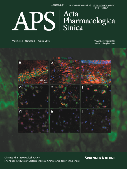
The expression of FSHR in depression-related brain regions. a The CA1 region of the mouse hippocampus. b The CA3 region of mouse hippocampus. c The DG of the mouse hippocampus. d The cortex. e The nucleus accumbens. f The amygdala. g The prefrontal cortex. h The bed nucleus of the stria terminalis. i The lateral habenula. FSHR (red), NeuN (green, a marker of neurons), and DAPI (blue) immunofluorescence was detected under a fluorescence microscope. See the article in pages 1033–1040.