Thank you for visiting nature.com. You are using a browser version with limited support for CSS. To obtain
the best experience, we recommend you use a more up to date browser (or turn off compatibility mode in
Internet Explorer). In the meantime, to ensure continued support, we are displaying the site without styles
and JavaScript.
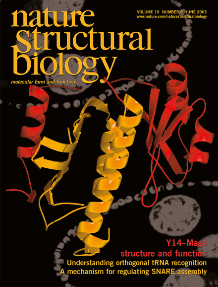
Crystal structure of the Drosophila protein Mago (yellow) in complex with Y14 (red). The complex is involved in post-splicing processes, such as nonsense-mediated mRNA decay and mRNA localization in Drosophila development. The background is a fluorescence microscope image showing co-localization of Y14 and Mago in the Drosophila egg chamber (Courtesy of O. Hachet and A. Ephrussi).
Expanding the genetic code by ribosome-mediated introduction of non-natural amino acids into proteins requires engineering of aminoacyl-tRNA synthetase–tRNA pairs. Structural information for pairs specific for tyrosine reveals how this goal may be achieved on a rational basis.
The notion that SNARE proteins constitute the minimal machinery for intracellular membrane fusion has been accepted by many scientists. An EPR study now brings new structural data on the neuronal SNARE VAMP-2 and emphasizes the importance of rigorous evidence before a hypothesis becomes a dogma.
Some ribosomal proteins recognize both ribosomal RNA and their own messenger RNA similarly. Although this phenomenon is widely conserved among prokaryotes for ribosomal protein S15, the precise manner in which the protein recognizes its own mRNA varies significantly from species to species.
Propagation of electrical signals by nerve cells relies on voltage-sensing ion channel proteins residing in the neuronal membrane. The first structure of a channel of this type dramatically overturns conventional wisdom about how these 'biomolecular transistors' work.