Volume 4
-
No. 12 December 2009
Cell bodies and proximal axons of rat sympathetic neurons on tracks in the proximal compartment of a 1-week-old compartmented (Campenot) culture. The neurons were fixed and stained for tubulin. Image is from the protocol by Campenot et al. Cover design by Jamel Wooten.
-
No. 11 November 2009
Vascular cast of choroidal vasculature taken with a scanning electron microscope. We thank Jolanta Sapieha for her expertise and technical help with the SEM image (Ecole Polytechnique, Montreal, Canada; supported by NSERC). Image is from the protocol by Connor et al. Cover design by Jamel Wooten.
-
No. 10 October 2009
Network constructed from mouse tissue atlas expression data. Nodes represent transcripts and edges correlations above r = 0.85. Each cluster of colored nodes represents a group of coexpressed genes. Image is from the protocol by Theocharidis et al. Cover design by Jamel Wooten.
-
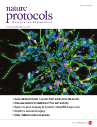
No. 9 September 2009
Spinal motor neurons (labeled by HB9 in red), along with other neurons (labeled by β III-tubulin in green), are differentiated from human embryonic stem cells using the stepwise protocol (all cell nuclei shown in blue). Image is from the protocol by Hu and Zhang. Cover design by Jamel Wooten.
-
No. 8 August 2009
Uropathogenic Escherichia coli bacteria exiting an infected mouse bladder superficial umbrella cell. Cover design by Jamel Wooten.
-
No. 7 July 2009
Adaptive reconstruction of the plasma membrane of a T cell from 3-D fluorescence microscopy image data based on iterative Voronoi optimization. Image is from the protocol by Klauschen et al. Cover design by Jamel Wooten.
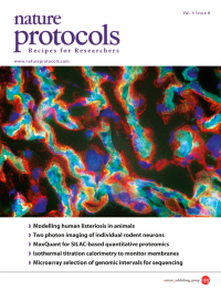
-
No. 6 June 2009
A 7 [µ]m immunolabeled gerbil placental tissue section showing co-expression of E-cadherin (red) and c-Met (green) in the syncytiotrophoblast barrier surrounding fetal capillaries (cyan). The proteins are receptors for Listeria monocytogenes invasion proteins InlA and InlB. Image is from the protocol by Disson et al. on p. 799. Cover design by Jamel Wooten.
-
No. 5 May 2009
DIC (differential interference contrast) image of a stained stage 9 Drosophila embryo hybridized with an RNA probe for the odd gene using a 96-well-plate high-throughput protocol. Image is from the protocol by Weiszmann et al. Photograph by A. Beaton. Cover design by Jamel Wooten.
-
No. 4 April 2009
Differential interference contrast images of stained Arabidopsis roots expressing an auxin responsive promoter fusion driving reporter expression. Image is from the protocol by Lewis and Muday on p. 437. Photographs were taken by Charles Buer and are reprinted with permission from Plant Cell (http://www.plantcell.org), copyright American Society of Plant Biologists, Plant Cell 16, 11911205 (2004).
-
No. 3 March 2009
Taking advantage of fluorophore-labeled oligonucleotides, the temperature-dependent assembly and disassembly of DNA nanostructures can be monitored in real time and with high throughput by FRET spectroscopy. Van't Hoff analysis of the fluorescence data provides insights into the thermodynamics of the self-assembly process. Image is from the protocol by Sacc[agrave] et al.
-
No. 2 February 2009
Multipotent adult germline stem cell (maGSC)-derived cardiac cluster stained with sarcomeric -actinin (green) and pan-cadherin (red). Image is from the protocol by Guan et al.
-
No. 1 January 2009
Calcispheres of Thoracosphaera heimii (recent sample top core) from the southeastern Indian Ocean. Image is from the protocol by Minoletti et al.