Volume 17 Issue 2, February 2022
Multiplexed imaging of diverse human tissues
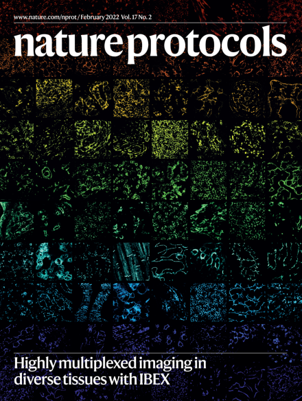
Rainbow collage of IBEX images from nine different human tissues, including the lymph node, thymus, spleen, jejunum, kidney, liver, skin and heart. Individual images display unique cell types and anatomical structures defined by a single protein biomarker. All protein biomarkers are targeted by commercially available antibodies and obtained by IBEX, an open-source multiplexed imaging method.
See Radtke et al.
Image: Andrea J. Radtke, CAT-I/LBS, LISB, NIAID, NIH. Cover design: Tulsi Voralia.