
Volume 6 Issue 9, September 2011
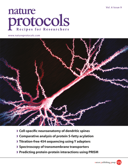
A monkey prefrontal layer III pyramidal cell injected with Lucifer yellow and imaged at high resolution on a confocal microscope. Final rendition is the product of tiling multiple 3D stacks. Image from the protocol by Dumitriu et al. (High throughput, detailed, cell-specific neuroanatomy of dendritic spines using microinjection and confocal microscopy. DOI: 10.1038/nprot.2011.389), in collaboration with J. Hao and TheVisualMD. Cover design by Jamel Wooten.
Protocol
-
Advertisement
