Thank you for visiting nature.com. You are using a browser version with limited support for CSS. To obtain
the best experience, we recommend you use a more up to date browser (or turn off compatibility mode in
Internet Explorer). In the meantime, to ensure continued support, we are displaying the site without styles
and JavaScript.
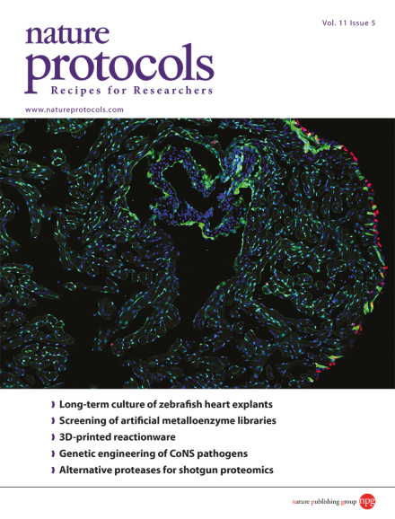
Cao and Poss (doi:10.1038/nprot.2016.049) describe methodology for culturing adult zebrafish heart explants and studying regeneration of epicardial tissue ex vivo. Shown is a section of a zebrafish heart explant that was cultured for 3 days. Endocardial and vascular endothelial cells are labeled green, cells that incorporated EdU in the previous day of culture are labeled red, and cell nuclei are labeled blue. Cover design by Jamel Wooten.
This protocol describes a genome-wide method to detect and to quantify DNA double-stranded breaks (DSBs). The approach is applicable to endogenous DSBs, but it can also be used to characterize the activity of engineered nucleases, including Cas9.
This protocol describes how to establish explant cultures of intact zebrafish hearts. The explants can be used to study the regeneration of cardiac tissues such as the epicardium, can be maintained for up to 30 d and are amenable to live imaging.
This protocol from Brautigam et al. describes methods for baseline correction and global analysis of isothermal titration calorimetry data using NITPIC and SEDPHAT. Publication-quality graphs of resulting data can then be created and visualized using GUSSI.
MicroED is a cryo-EM technique for collecting electron diffraction data from microcrystals and nanocrystals. This protocol from Gonen and colleagues describes how to prepare the protein crystal samples, how to set up the electron microscope for MicroED, and diffraction data collection.
This protocol describes the use of the AutoDock suite for computational docking in the study of protein–ligand interactions. A number of methods are described ranging from basic docking of drug molecules to virtual screening using a large ligand library of chemical compounds.
This protocol describes a rapid (<24 h), reproducible and cheap technique for evaluating cancer cell invasiveness by quantifying in vivo rates of cancer cell extravasation in the chorioallantoic membrane (CAM) of chicken embryos.
This protocol provides a method for genetically engineering untransformable coagulase-negative staphylococci (CoNS), using bacteriophage Φ187–mediated plasmid transfer. This is a major technical advancement that enables research on CoNS-mediated infections.
Here the authors provide the procedural details of a new approach to clone large (up to 100 kb) microbial genomic sequences using Cas9-assisted targeting of chromosome segments (CATCH). The Cas9 cleavage is achieved in agarose gel, and the cleaved product can be ligated into a cloning vector.
This protocol for the induction and isolation of Plasmodium falciparum gametocytes combines seven parameters that have been shown to facilitate the optimum induction of gametocytogenesis in vitro to obtain highly synchronous gametocyte stages on a large scale.
The use of a single enzyme such as trypsin for shotgun proteomics limits the ability to cover the whole proteome and all protein post-translational modifications. This protocol describes the use of six alternative proteases that complement trypsin to increase the coverage of the proteome.