Thank you for visiting nature.com. You are using a browser version with limited support for CSS. To obtain
the best experience, we recommend you use a more up to date browser (or turn off compatibility mode in
Internet Explorer). In the meantime, to ensure continued support, we are displaying the site without styles
and JavaScript.
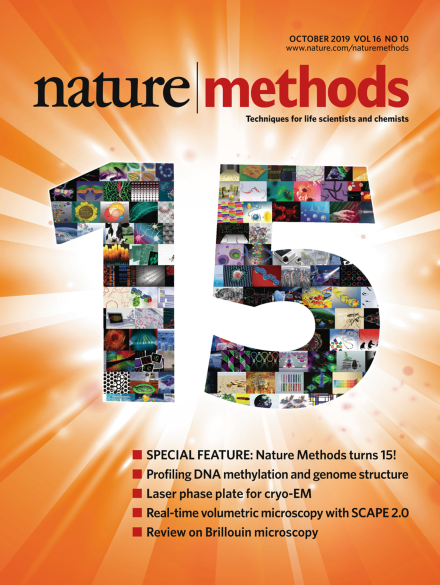
This month we celebrate the fifteen-year anniversary of Nature Methods. The cover artwork comprises images from previous covers of Nature Methods throughout the years.
We celebrate the 15th anniversary issue of Nature Methods with a look back on the past decade and a half, and a look forward to the future of methods development.
Single-particle techniques offer an unprecedented opportunity to understand the role of structural variability in biological function. They also call into question the meaning of ‘a structure’ and its relevance to function.
To mark the 15th anniversary of Nature Methods, we asked scientists from across diverse fields of basic biology research for their views on the most exciting and essential methodological challenges that their communities are poised to tackle in the near future.
Labs in different neuropsychiatry subfields don’t always see eye to eye, but convergent approaches help them join forces to study these difficult conditions.
This Review discusses the principles, advantages and limitations of Brillouin microscopy, a non-invasive tool for measuring mechanical properties of biological samples in three dimensions.
Garnett uses a hierarchical markup language and machine learning to define cell types and their marker genes and identifies these cell types in scRNA-seq datasets from tissues and whole organisms and across species.
Methyl-HiC combines the elucidation of chromatin architecture with the reading of DNA methylomes in pools and single cells. Regions that are distant on the linear-genome but close in three-dimensional space show coordinated DNA methylation.
Functional ultrasound (fUS) imaging of neural activity has been extended to volumetric imaging across the whole brain. 4D fUS is demonstrated in the rat brain in response to sensory stimuli and during seizure-like activity.
Single-nucleus methyl-3C sequencing jointly interrogates 3D chromatin organization and DNA methylation in human cells, and these joint measurements more accurately distinguish different cell types than either unimodal method.
CellAssign uses a probabilistic model to assign single cells to a given cell type defined by known marker genes, enabling automated annotation of cell types present in a tumor microenvironment.
Phase plates improve contrast in cryo-electron microscopy, but suffer from electrostatic charging and electron scattering. A laser phase plate overcomes these problems and may improve imaging of biological specimens.
Mass spectrometry imaging with fluoromethylpyridinium-based reactive matrices allows mapping of neurotransmitters at high resolution and at a low limit of detection. The approach is applied to rat, macaque and human brain tissue samples for probing Parkinson’s disease-related changes.
Fusion to fluorescent proteins enables efficient two-photon activation of blue-light-controlled optical dimerizers via FRET. FRET-assisted photoactivation was used to study extracellular signal-regulated kinase activation in 3D epithelial cysts, organoids and living mice.
A computational workflow combining image segmentation, computer graphics and supervised machine learning enables automated and robust 3D analysis of the coupling of cell shape and signaling.
Cell lines in which Nup96 is endogenously tagged with mEGFP, SNAP-tag, HaloTag or mMaple serve as versatile reference samples, enabling 3D resolution calibration, assessment of labeling efficiency and precise molecular counting.
SCAPE 2.0 is a versatile imaging platform that enables real-time three-dimensional microscopy of cellular function and dynamic motion in living organisms at over 100 volumes per second with minimal photodamage, and high-throughput structural imaging in fixed, cleared and expanded samples.