Thank you for visiting nature.com. You are using a browser version with limited support for CSS. To obtain
the best experience, we recommend you use a more up to date browser (or turn off compatibility mode in
Internet Explorer). In the meantime, to ensure continued support, we are displaying the site without styles
and JavaScript.
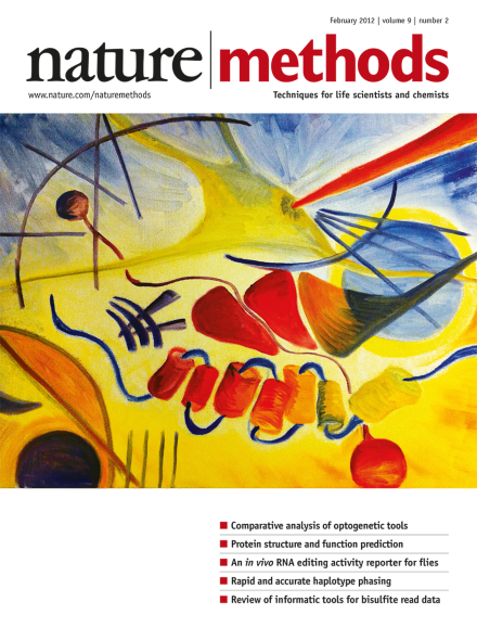
Image of an original oil painting by Emily Ferenczi–a graduate student in the laboratory of Karl Deisseroth–inspired by Wassily Kandinsky's Yellow-Red-Blue. Although individual elements are open to interpretation, this painting depicts an experiment involving optogenetic control of neuronal activity. Analysis p159
Direct comparisons of tool or method performance under standardized experimental conditions yield highly valuable information for both method users and developers.
The use of a photoswitchable fluorescent protein allows nonlinear structured-illumination microscopy of cellular structures at 50 nanometer resolution.
A sophisticated analysis approach based on the concept of fluorophore localization provides dynamic super-resolution data of GFP-labeled live cells using a common, arc lamp–based wide-field fluorescence microscope.
In vivo methods to capture processing events such as RNA editing in specific cell types are sparse. Researchers have now developed a method to visualize adenosine-to-inosine editing activity in individual fruit fly neurons using a reverse-engineered fluorescent reporter.
In this Review, the authors take the reader through the steps needed to analyze bisulfite-treated DNA, pointing out different considerations for data in base or color space, to ensure high-quality methylome analysis.
In this Perspective the authors highlight and discuss the artifacts that can arise when using immunolabeling to examine protein localization in cell culture. They call for using both alternative fixation and permeabilization protocols and live-cell imaging of fluorescent protein fusions to reliably study subcellular protein localization.
In this Analysis, the authors directly experimentally compare microbial opsins used for the control of neural activity. They extract essential principles and key parameters that can help end users with the design and interpretation of optogenetic experiments and guide tool developers in the characterization of future tools.
HHblits is a protein sequence search tool that works by iterative pairwise comparison of profile hidden Markov models. It outperforms existing tools in terms of speed, sensitivity and alignment quality.
The Splitread algorithm uses a split-read strategy to detect structural variants and small insertions and deletions (indels) in whole-exome and whole-genome sequence datasets at high sensitivity. It maps the breakpoints at single-base-pair resolution, even in low-complexity regions, and can detect novel processed pseudogenes.
An efficient haplotype-estimation algorithm that features linear complexity allows the rapid and accurate phasing of diploid genomes from trios, duos and unrelated samples.
A simple, general procedure for transferring protein complexes directly from native gels to electron microscopy grids for structural analysis is reported.
The use of dual-objective detection with astigmatism-based three-dimensional stochastic optical reconstruction microscopy (STORM) imaging improves resolution more than twofold and removes noise in resulting super-resolution images. This allowed detailed fluorescence imaging of distinctive features of the three-dimensional actin cytoskeleton ultrastructure with single-filament resolution in cells.
Adenosine-to-inosine RNA editing modifies expressed sequences and enhances functional protein diversity. The authors report an in vivo fluorescent reporter that provides a readout of adenosine deaminase RNA-editing activity in Drosophila melanogaster neurons, showing evidence of inter-individual variability in editing activity.
Bayesian analysis of fluorophore blinking and bleaching in image data collected from simple xenon arc lamp illumination and high-speed wide-field imaging of standard fluorescent proteins allows localization microscopy in living cells with a 50 nm spatial and a 4 s temporal resolution.
An acousto-optic two-photon microscope with continuous three-dimensional trajectory and random-access scanning modes can scan near-cubic-millimeter volumes of tissue at sub-millisecond temporal resolution in vivo. The system can be used to image both sub-cellular as well as network-scale neuronal activity.