Thank you for visiting nature.com. You are using a browser version with limited support for CSS. To obtain
the best experience, we recommend you use a more up to date browser (or turn off compatibility mode in
Internet Explorer). In the meantime, to ensure continued support, we are displaying the site without styles
and JavaScript.
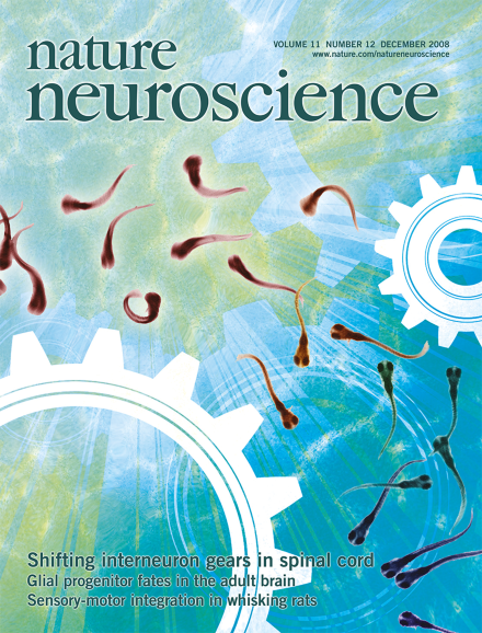
Increasingly forceful movements are thought to arise from the recruitment of additional cells to the active motoneuron pool. McLean and colleagues now demonstrate that two completely different classes of spinal premotor interneurons drive motoneurons during slow and fast swimming of zebrafish larvae. As the fish accelerate, the 'slow' interneurons are progressively silenced while the 'fast' interneurons take over.
Glial cells that express NG2 and platelet-derived growth factor receptor α are found throughout the mature CNS. These cells are mitotically active, but their functions remain enigmatic. A genetic fate-mapping study in this issue shows that these abundant glial cells can generate both oligodendrocytes and some cortical projection neurons in the adult brain.
A study in this issue shows that zebrafish larvae deploy different groups of excitatory spinal interneurons to drive slow, fast and top speed swimming. As one set is gradually activated, the others are partially or fully inhibited.
Demonstrating how specific motor signals modulate sensory processing in the rat vibrissal system, a new study in this issue shows that motor signals first attenuate and then amplify afferent sensory signals.
Correlations in firing rate between pairs of neurons can change depending on task and attentional demands. This new finding suggests that measuring correlations can help to reveal how neural circuits process information.
Following brief stimulation, macroscopic NMDA receptor currents decay with biphasic kinetics believed to reflect glutamate dissociation and receptor desensitization. The authors show that the fast and slow decay components arise from the deactivation of receptor populations that gate with short and long openings.
fMRI studies suggest that nucleus accumbens (NAc) activation increases in response to stimuli of different hedonic valence, whereas physiological evidence suggests that NAc neurons show increases in activity for rewarding stimuli and pauses for aversive stimuli. Using cyclic voltammetry, the authors find that patterns of dopamine release and metabolic activity differentiate between rewarding and aversive stimuli.
No two roses smell exactly alike, but our brain accurately bundles these variations into a single percept 'rose'. The authors now report that although olfactory bulb neurons decorrelate odor mixtures that are quite similar, piriform cortex neuronal responses show pattern completion and predict olfactory perception.
Dopamine is known to contribute to the amygdala-mediated aversive response, where increased dopamine release can augment amygdala function. Combining fMRI and PET imaging techniques, Kienast et al. present findings that suggest a functional link between anxiety temperament, dopamine storage capacity and emotional processing in the amygdala.
Genetic ablation of β-catenin in the embryonic ventral forebrain restricted proliferation of neural precursors in the medial ganglionic eminence, resulting in fewer cholinergic projection neurons in basal forebrain and fewer calbindin- and somatostatin-positive interneurons in the cortex. This work suggests a crucial role for canonical Wnt signaling in ventral forebrain neurogenesis.
About 4% of the cells in the adult rodent brain are PDGFRA+ NG2+ glia, derived from the oligodendrocyte lineage. Rivers and colleagues constructed a transgenic mouse to fate map the PDGFRA+ glia. In the adult corpus callosum, these cells generated substantial numbers of late-myelinating oligodendrocytes. In the cortex, little late myelination was observed; instead, PDGFRA+ precursors seemed to continuously generate small numbers of projection neurons mainly in piriform cortex.
Adenosine receptor A2AR is known to antagonize dopaminergic signaling in the striatum and its malfunctions have been implicated in various striatum-related diseases. Flajolet et al. show that A2AR and fibroblast growth factor receptor interact to synergistically activate ERK1/2 pathway and that such interaction modulates the morphological changes of cultured neurons and synaptic plasticity of cortico-striatal synapses.
Hippocampal GABAergic synapses are excitatory during the early postnatal period and can undergo spike timing-dependent modifications of synaptic strength. Xu and colleagues demonstrate that this plasticity can be modulated bidirectionally by frequency and that it depends on the action of GABAB receptors.
The authors here show that two completely different classes of spinal premotor interneurons drive motoneurons during slow and fast swimming of zebrafish larvae. As the fish accelerate, the 'slow' interneurons are progressively silenced, while the 'fast' interneurons take over, and vice versa.
In sedated and whisking rats, the authors show that motor cortex activity enhances sensory processing through a cortico-cortico-thalamic feedback circuit. In whisking rats, however, inhibitory brainstem input to the thalamus was also enhanced, leading to a net suppression of thalamic sensory responses.
The authors report that fMRI responses in human foveal retinotopic cortex contain information about objects presented in the periphery. This information is position invariant and correlated with perceptual discrimination accuracy.
It has been proposed that neurons in the intraparietal cortex gradually accumulate evidence supporting different response options. Here the authors show that this model generalizes to arbitrary stimulus-response associations in humans.
The nervous system produces accurate movements by adapting to environmental changes. The authors construct a probabilistic model that compensates for motor errors and estimates their sources, finding that if the motor system used such a strategy, it would explain many previously observed movement-generalization phenomena.