


« Prev Next »

Cell Membranes
Cell membranes protect and organize cells. All cells have an outer plasma membrane that regulates not only what enters the cell, but also how much of any given substance comes in. Unlike prokaryotes, eukaryotic cells also possess internal membranes that encase their organelles and control the exchange of essential cell components. Both types of membranes have a specialized structure that facilitates their gatekeeping function.
What Are Cellular Membranes Made Of?
With few exceptions, cellular membranes — including plasma membranes and internal membranes — are made of glycerophospholipids, molecules composed of glycerol, a phosphate group, and two fatty acid chains. Glycerol is a three-carbon molecule that functions as the backbone of these membrane lipids. Within an individual glycerophospholipid, fatty acids are attached to the first and second carbons, and the phosphate group is attached to the third carbon of the glycerol backbone. Variable head groups are attached to the phosphate. Space-filling models of these molecules reveal their cylindrical shape, a geometry that allows glycerophospholipids to align side-by-side to form broad sheets (Figure 1).
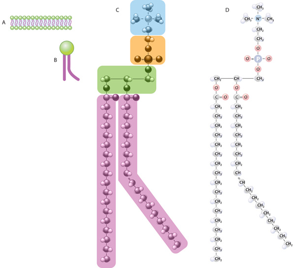
Figure 1: The lipid bilayer and the structure and composition of a glycerophospholipid molecule
(A) The plasma membrane of a cell is a bilayer of glycerophospholipid molecules. (B) A single glycerophospholipid molecule is composed of two major regions: a hydrophilic head (green) and hydrophobic tails (purple). (C) The subregions of a glycerophospholipid molecule; phosphatidylcholine is shown as an example. The hydrophilic head is composed of a choline structure (blue) and a phosphate (orange). This head is connected to a glycerol (green) with two hydrophobic tails (purple) called fatty acids. (D) This view shows the specific atoms within the various subregions of the phosphatidylcholine molecule. Note that a double bond between two of the carbon atoms in one of the hydrocarbon (fatty acid) tails causes a slight kink on this molecule, so it appears bent.
© 2010 Nature Education All rights reserved. 
Glycerophospholipids are by far the most abundant lipids in cell membranes. Like all lipids, they are insoluble in water, but their unique geometry causes them to aggregate into bilayers without any energy input. This is because they are two-faced molecules, with hydrophilic (water-loving) phosphate heads and hydrophobic (water-fearing) hydrocarbon tails of fatty acids. In water, these molecules spontaneously align — with their heads facing outward and their tails lining up in the bilayer's interior. Thus, the hydrophilic heads of the glycerophospholipids in a cell's plasma membrane face both the water-based cytoplasm and the exterior of the cell.
Altogether, lipids account for about half the mass of cell membranes. Cholesterol molecules, although less abundant than glycerophospholipids, account for about 20 percent of the lipids in animal cell plasma membranes. However, cholesterol is not present in bacterial membranes or mitochondrial membranes. Also, cholesterol helps regulate the stiffness of membranes, while other less prominent lipids play roles in cell signaling and cell recognition.
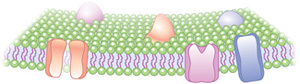
Figure 2: The glycerophospholipid bilayer with embedded transmembrane proteins
© 2010 Nature Education All rights reserved.
At physiological temperatures, cell membranes are fluid; at cooler temperatures, they become gel-like. Scientists who model membrane structure and dynamics describe the membrane as a fluid mosaic in which transmembrane proteins can move laterally in the lipid bilayer. Therefore, the collection of lipids and proteins that make up a cellular membrane relies on natural biophysical properties to form and function. In living cells, however, many proteins are not free to move. They are often anchored in place within the membrane by tethers to proteins outside the cell, cytoskeletal elements inside the cell, or both.
What Do Membranes Do?
Cell membranes serve as barriers and gatekeepers. They are semi-permeable, which means that some molecules can diffuse across the lipid bilayer but others cannot. Small hydrophobic molecules and gases like oxygen and carbon dioxide cross membranes rapidly. Small polar molecules, such as water and ethanol, can also pass through membranes, but they do so more slowly. On the other hand, cell membranes restrict diffusion of highly charged molecules, such as ions, and large molecules, such as sugars and amino acids. The passage of these molecules relies on specific transport proteins embedded in the membrane.
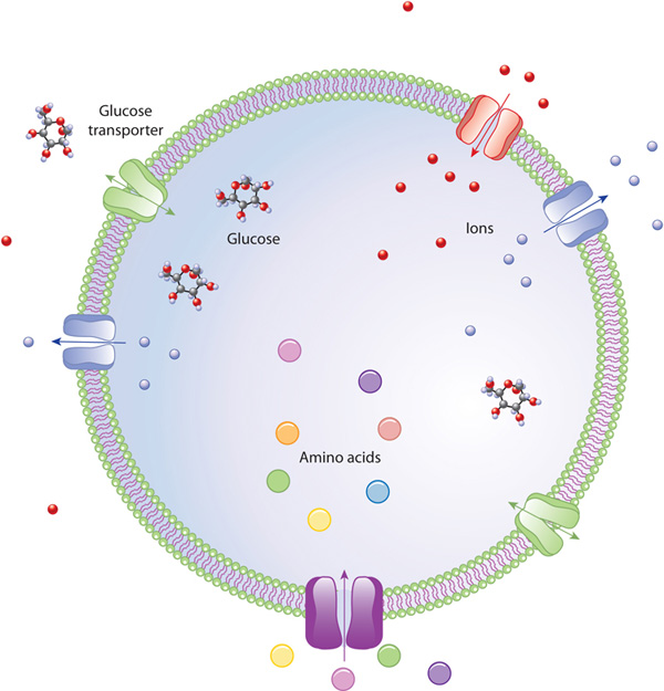
Figure 3: Selective transport
Specialized proteins in the cell membrane regulate the concentration of specific molecules inside the cell.
© 2010 Nature Education All rights reserved.
Membrane transport proteins are specific and selective for the molecules they move, and they often use energy to catalyze passage. Also, these proteins transport some nutrients against the concentration gradient, which requires additional energy. The ability to maintain concentration gradients and sometimes move materials against them is vital to cell health and maintenance. Thanks to membrane barriers and transport proteins, the cell can accumulate nutrients in higher concentrations than exist in the environment and, conversely, dispose of waste products (Figure 3).
Other transmembrane proteins have communication-related jobs. These proteins bind signals, such as hormones or immune mediators, to their extracellular portions. Binding causes a conformational change in the protein that transmits a signal to intracellular messenger molecules. Like transport proteins, receptor proteins are specific and selective for the molecules they bind (Figure 4).
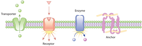
Figure 4: Examples of the action of transmembrane proteins
Transporters carry a molecule (such as glucose) from one side of the plasma membrane to the other. Receptors can bind an extracellular molecule (triangle), and this activates an intracellular process. Enzymes in the membrane can do the same thing they do in the cytoplasm of a cell: transform a molecule into another form. Anchor proteins can physically link intracellular structures with extracellular structures.
© 2010 Nature Education All rights reserved.
Peripheral membrane proteins are associated with the membrane but are not inserted into the bilayer. Rather, they are usually bound to other proteins in the membrane. Some peripheral proteins form a filamentous network just under the membrane that provides attachment sites for transmembrane proteins. Other peripheral proteins are secreted by the cell and form an extracellular matrix that functions in cell recognition.
How Diverse Are Cell Membranes?
In contrast to prokaryotes, eukaryotic cells have not only a plasma membrane that encases the entire cell, but also intracellular membranes that surround various organelles. In such cells, the plasma membrane is part of an extensive endomembrane system that includes the endoplasmic reticulum (ER), the nuclear membrane, the Golgi apparatus, and lysosomes. Membrane components are exchanged throughout the endomembrane system in an organized fashion. For instance, the membranes of the ER and the Golgi apparatus have different compositions, and the proteins that are found in these membranes contain sorting signals, which are like molecular zip codes that specify their final destination.
Mitochondria and chloroplasts are also surrounded by membranes, but they have unusual membrane structures — specifically, each of these organelles has two surrounding membranes instead of just one. The outer membrane of mitochondria and chloroplasts has pores that allow small molecules to pass easily. The inner membrane is loaded with the proteins that make up the electron transport chain and help generate energy for the cell. The double membrane enclosures of mitochondria and chloroplasts are similar to certain modern-day prokaryotes and are thought to reflect these organelles' evolutionary origins.
Conclusion
Membranes are made of lipids and proteins, and they
serve a variety of barrier functions for cells and intracellular
organelles.
Membranes keep the outside "out" and the inside "in," allowing only
certain
molecules to cross and relaying messages via a chain of molecular events
eBooks
This page appears in the following eBook


