Volume 36
-
No. 13 December 2011
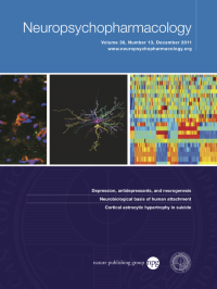
Patterns of activation-signal recorded from the right amygdala in mothers while presented with own and unfamiliar infant-related video vignettes. Signal patterns were measured for two groups of mothers on the basis of micro-coding maternal and infant social behavior during mother-infant interaction: synchronous mothers (S—orange)—those who coordinate social behavior with infant signals- and intrusive mothers (I—red) —those providing access stimulation when infants signal a need for rest. As seen, among synchronous mothers, activations of the right amygdala show greater coherence over time, whereas among intrusive mothers activation of this nucleus show greater disorganization. Courtesy of the authors from the Department of Psychology, Bar-Ilan University, Israel and the Functional brain center, Wohl institute for advanced imaging, Tel-Aviv Sourasky medical center: Shir Atzil, Talma Hendler and Ruth Feldman.
Supplement
-
No. 12 November 2011
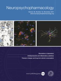
3-D reconstruction of a representative white matter astrocyte in the anterior cingulate cortex of depressed suicides (courtesy of Dr Naguib Mechawar, McGill Group for Suicide Studies, McGill University, Canada).
-
No. 11 October 2011
One micron optical confocal sections through the amygdala region of a mouse transgenic for the GAL5.1hβg-lacZ transgene following fluorescent immunohistochemistry using antisera against the galanin peptide (false colour green) and β-galactosidase (false colour red). Colocalisation is highlighted in yellow. Courtesy of the authors from the School of Medical Sciences, University of Aberdeen, Scotland: Scott Davidson, Marissa Lear, Lynne Shanley, Benjamin Hing, Amanda Baizan-Edge and Alasdair MacKenzie; the authors from Rowett Institute of Nutrition and Health, Greenburn Aberdeen, Scotland: Annika Herwig & Perry Barrett; the author from the School of Biomedical Sciences, University of Liverpool: John Quinn; the authors from Institute of Psychiatry, King's College London, UK: Gerome Breen & Peter McGuffin; and the author from School of Engineering, Kings College, University of Aberdeen, Scotland: Andrew Starkey.
-
No. 10 September 2011
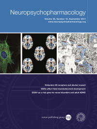
An electron micrograph of the rat prefrontal cortex, with asymmetric (excitatory) synapses (orange) identified between dendritic spines (blue) and boutons by their characteristically electron dense postsynaptic zone. Following treatment with phencyclidine, there is a persistent decrease in number of asymmetric synapses and dopamine tone in prefrontal cortex, changes that are hypothesized to underlie the cognitive deficit in this animal model of schizophrenia. This spine synapse loss in prefrontal cortex is reversed by acute treatment with the atypical antipsychotic drug, olanzapine, and this reversal is maintained by chronic oral treatment, paralleling the time course of the restoration of the dopamine deficit, and normalization of cognitive function produced by olanzapine. Courtesy of Drs. John Elsworth, Tibor Hajszan, Csaba Leranth and Robert Roth, Yale University, USA.
-
No. 9 August 2011
Nicotine lozenges enhance cognitive brain activity during smoking withdrawal, as assessed with simultaneous functional MRI and electroencephalography (EEG). This figure shows regions of the brain where cognition-related fMRI activation (orange/yellow) and deactivation (blue) were inversely associated with scalp electrophysiological activity in the alpha frequency range. Courtesy of Dr John Beaver, GSK Clinical Imaging Centre and Imperial College, London, UK.
-
No. 8 July 2011
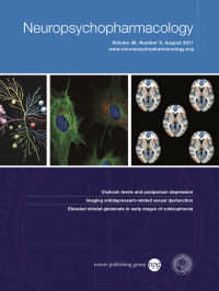
Immuno-reactivity of glial fibrillary acidic protein (green) in primary astrocytes treated with amitriptyline. Lysosomes are stained with an acid-dependent lysotracker dye (red). Courtesy of Ms. Vanessa Ganal, Drs. Jürgen Zschocke and Theo Rein, MPI of Psychiatry, Germany.
-
No. 7 June 2011
Offered by scientific illustrator Katie Vicari of Nature Publishing, this figure is an artistic adaptation based on KINOMEscan profiling technology (DiscoveRx, CA) and kinase dendrogram image (Cell Signaling Technology, MA). The compound was screened against 359 non mutant human kinases for competitive binding with active site binders. Competitive binding relative to positive controls (% of control) is shown, with larger spheres indicating higher binding affinity. Original figure is courtesy of Dr Jen Pan et al, of Broad Institute of Harvard and MIT, Cambridge, MA.
-
No. 6 May 2011
Pattern of brain activity that differentiates methylphenidate (red) from atomoxetine (blue) during the delay component of a rewarded working memory task. Courtesy of the Department of Neuroimaging, Institute of Psychiatry, King's College, London, UK (corresponding author: Andre Marquand, andre.marquand@kcl.ac.uk).
-
No. 5 April 2011
Calphostin C treatment reduces the level of β-catenin in SH-SY5Y cells transfected with APPsw. Courtesy of Professor Zhan-You Wang, et al Key Laboratory of Medical Cell Biology of Ministry of Education, and Key Laboratory of Endocrine Diseases of Liaoning Province, China Medical University, Shenyang, China.
-
No. 4 March 2011
Illustration of tract-based quantification of fractional anisotropy. The labeling of genu of corpus callosum (yellow) is overlaid on the skeleton (green) of the white matter to measure the fractional anisotropy change of this tract. Courtesy of Drs. Hao Huang and Uma Rao, University of Texas Southwestern Medical Center.
-
No. 3 February 2011
Triple-label immunostaining in the PAG. There was an almost complete colocalization between GFAP and TNFα imaging, but TNFα did not colocalize with NeuN, which suggested that TNFa was located on astrocytes, but not neurons; courtesy of Dr. Shuanglin Hao, Department of Anesthesiology, University of Miami, FL.
-
No. 2 January 2011
Firing of neurons in the nucleus accumbens synchronizes best to ventral hippocampus of the past, as measured by the phase locking of accumbens neurons to theta-frequency activity in the hippocampus. See Figure 6, Nason et al, page 494, courtesy of MW Nason, SUNY Stony Brook, Stony Brook, NY, USA.