Thank you for visiting nature.com. You are using a browser version with limited support for CSS. To obtain
the best experience, we recommend you use a more up to date browser (or turn off compatibility mode in
Internet Explorer). In the meantime, to ensure continued support, we are displaying the site without styles
and JavaScript.
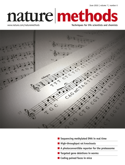
A DNA sequence put to music. A, C and G are represented by the notes of the same name. For T, the solfege syllable 'ti' is used, which is E in the key of F major (chosen because it includes the other three pitches as well). All of the notes are quarter notes except for the methyl C (represented by a half note with a fermata) and notes in the vicinity of a methyl C, where dotted quarters and eighth notes create syncopation and rhythmic variation. For a trumpet rendition of the piece, please visit Methagora. Cover idea courtesy of Pacific Biosciences; cover design by Erin Dewalt. Article p461
New genetic technologies in the rat open new research opportunities and call for more careful choice of the most suitable model organism before embarking on a study.
High-throughput imaging of genome-wide RNA interference screens and systematic analysis of protein complexes involved in mitosis yield valuable resources and reveal new subunits of well-characterized complexes.
Applying technology developed for next-generation DNA sequencing to study translation, researchers watch individual ribosomes string together amino acids in real time.
A new technique for transcriptome-wide isolation of RNAs bound to specific proteins reveals, with high resolution, the location of RNA-binding proteins on their target RNAs.
The Dynameomics database houses native-state and high-temperature unfolding simulation data for the 100 most populated protein folds; such information provides insights into protein function beyond structure alone.
Researchers try multiple means to get high-quality membrane proteins for X-ray crystallography and nuclear magnetic resonance (NMR) spectroscopy studies.
Measuring the kinetics of nucleotide incorporation during single-molecule, real-time DNA sequencing allows identification of methylated bases during the sequencing process.
The arsenal of methods to investigate gene function in Caenorhabditis elegans continues to grow—with new approaches to generate targeted deletion mutants and to control gene expression.
Cultured rat spermatogonia, genetically modified via a transposon-based gene trap, are inserted into the testes of sterile rats and give rise to mutant progeny with many different genotypes.
Targeted deletion of genes within 25 kb of Mos1 transposons in the C. elegans genome is demonstrated. This should enable single-gene deletions of more than 8,000 genes for which deletions are currently not available in this organism.
This computational process evaluates gene models in prokaryotic genomes, independently of the gene finder used, and reports anomalies that can be used to improve the quality of gene models through manual curation.
The activity of zinc-finger nucleases in mammalian cells is enhanced by transient incubation of the cells at low temperatures. Incorporation of this simple step should improve the efficiency of these tools for genome manipulation.
Polymerase kinetics observed during single-molecule, real-time sequencing depend on the methylation status of the DNA template. Measurement of kinetic parameters such as interpulse duration and pulse width allows the identification of methylated adenosine in Escherichia coli and the distinction between 5-methylcytosine and 5-hydroxymethylcytosine in synthetic templates.
Combining reverse transfection of protein tyrosine kinase substrates on cell arrays with fluorescence resonance energy transfer (FRET) measurements by fluorescence lifetime imaging microscopy (FLIM) allows quantitative assessment of phosphorylation patterns and identification of feedback loops at single-cell resolution.
A photoconvertible reporter of the ubiquitin-proteasome system permits detection of its activity independent of protein synthesis and is applied to study cell type– and age-specific protein degradation in living Caenorhabditis elegans.