Thank you for visiting nature.com. You are using a browser version with limited support for CSS. To obtain
the best experience, we recommend you use a more up to date browser (or turn off compatibility mode in
Internet Explorer). In the meantime, to ensure continued support, we are displaying the site without styles
and JavaScript.
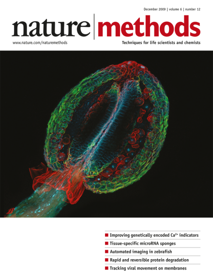
This photograph of an Arabidopsis thaliana (thale cress) anther took first place in the 2009 Nikon Small World photomicrography competition. The image was taken by Heiti Paves of Tallinn University of Technology in Tallinn, Estonia using a confocal microscope at ×20 magnification. Other images from this year's competition are on display at http://www.nikonsmallworld.com/.
Neuroscience methods are undergoing a dramatic change owing to improvements in optical probes, but standardized evaluation procedures would aid probe development and uptake.
Two methods enable the drawing of genome-wide chromatin interaction maps: one looks at protein-independent folding principles, the other at protein-mediated functional interactions.
Researchers observe that cells of the post-implantation mouse epiblast can revert to an embryonic stem cell–like state without the addition of exogenous genes.
An improved version of the GCaMP genetically encoded calcium indicator, called GCaMP3, has higher calcium affinity and increased baseline fluorescence, dynamic range and stability. GCaMP3 performs better than existing genetically encoded calcium indicators in several assays and organisms, including in vivo imaging of neuronal signaling in worms, flies and mice.
Fusion of the genetically-encoded calcium indicator GCaMP2 to synaptophysin localizes the sensor to neuron presynaptic terminals and conveys linear responsiveness over a wider range of spike frequencies. The sensor allowed measurement of synaptic activity caused by spiking as well as graded voltage signals during in vivo imaging in zebrafish.
Neuronal stimulation with channelrhodopsin-2 is combined with calcium fluorescence imaging to study neural connections in intact Caenorhabditis elegans.
Tissue-specific expression of microRNA sponges allows precise regulation of microRNA activity in living flies. The authors investigate the role of miR-8 in the formation of neuromuscular junctions in detail.
Microsources positioned with holographic optical tweezers can establish a highly localized, three-dimensional chemical gradient that allows the manipulation of polarization and migration in single cells.
Methods for automated fluorescence imaging allow high-throughput examination of reporter expression patterns in zebrafish embryos. They are applied to mapping promoter-enhancer interactions in this organism.
A degradation pathway found in plants, dependent on the hormone auxin, can be transplanted and harnessed to induce rapid and reversible target protein degradation in both yeast and animal cells.
A combination of scattering interferometry and single-molecule fluorescence microscopy allows visualization of both the position and orientation of single Simian virus 40 particles on lipid bilayers and provides evidence of viral interaction with receptors in membrane nanodomains.
They are the quintessential drug target—but the dynamic structures and highly elaborate mechanisms of G protein–coupled receptors continue to keep experts in both industry and academia on their toes.