Volume 10
-
No. 12 December 2013
Releasing intact membrane protein complexes in bicelles or nanodiscs into the gas phase for observation by mass spectrometry. Photograph and cover art by Jonathan Hopper, Karl Harrison and Michelle Smikle. Brief Communication p1206
-
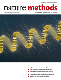
No. 11 November 2013
Chaetoceros debilis, a colonial diatom captured from marine plankton in the North Sea and photographed using differential interference contrast microscopy by Wim van Egmond of the Micropolitan Museum, The Netherlands. Winner of the 2013 Nikon Small World photomicrography contest (reprinted with permission from Nikon).
-
No. 10 October 2013
A collection of human knockout cell lines. Cover by Erin Dewalt, based on a concept by Nicola Graf (freelance designer) and image from iStockphoto/Thinkstock. Resource p965
-
No. 9 September 2013
One-dimensional superresolution imaging of individual proteins bound to densely covered DNA that is stretched between two optically trapped microspheres. Image by Ivo van der Ent (Vrije Universiteit Amsterdam). Article p910.
-
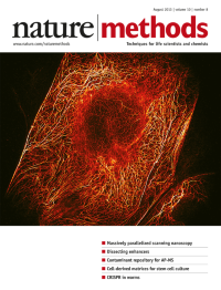
No. 8 August 2013
RESOLFT image of keratin filaments in kidney epithelial cells. The total image acquisition time was ~2 s. Image by Andriy Chmyrov and Stefan Hell. Brief Communication p737
-
No. 7 July 2013
DNA on a pH-sensing semiconductor microchip. Image by Ken Yang (freelance designer). Article p641
-
No. 6 June 2013
Visualization of functional connectivity in the human cerebral cortex based on magnetic resonance imaging data. Brain image by Joachim Böttger and Daniel Margulies (Max Planck Institute for Human Cognitive and Brain Sciences, Leipzig, Germany) with compositing by Tobias S. Hoffmann. Cover composition by Erin Dewalt. Focus p479
-
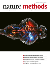
No. 5 May 2013
Bursts of neural activity in an entire larval zebrafish brain expressing a calcium reporter in every neuron. Two different time points are shown in red and blue. Image by Philipp Keller, Misha Ahrens and Kristin Branson (Howard Hughes Medical Institute, Janelia Farm Research Campus).
-
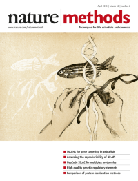
No. 4 April 2013
The cover is an artistic depiction of genome engineering in the zebrafish. Illustration by Vivian Lin, Harvard-Westlake School. Cover design by Erin Dewalt.
-
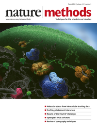
No. 3 March 2013
Hfq hexamers in different binding states with different mobilities in the intracellular environment. Cover image by the Elf lab, Irmeli Barkfors and Tremani. Article p265
-
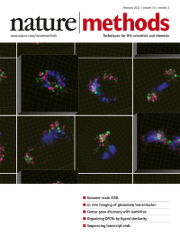
No. 2 February 2013
Chromosome territories in human cells are shown in these three-dimensional renderings of high-definition fluorescence in situ hybridization images. Cover by Erin Dewalt, based on images by Magda Bienko and Alexander van Oudenaarden. Article p122
-
No. 1 January 2013
Mass spectrometry–based targeted proteomics, our choice for Method of the Year 2012, is allowing biologists to follow sets of proteins with high sensitivity, reliability and quantitative accuracy. Cover design by Erin Dewalt. Special feature starts on p19.