Volume 9
-
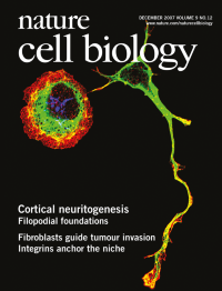
No. 12 December 2007
A fluorescence image of two cortical neurons labelled for filamentous actin (red) and microtubules (green). The round cell on the left was transfected with a construct that sequesters Ena/VASP proteins to mitochondria (blue); this inhibits filopodia formation and neurite outgrowth.
-
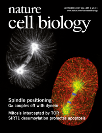
No. 11 November 2007
During anaphase of one-cell stage Caenorhabditis elegans embryos, spindle positioning is mediated by microtubules (white and green), as well as dynein (red).
-
No. 10 October 2007
Post-mitotic nuclear envelope assembly involves ER reorganization by chromatin.
-
No. 9 September 2007
This issue of Nature Cell Biology includes a series of six specially commissioned articles that collectively highlight how misfunctioning developmental pathways can cause disease. The articles are freely accessible until February 2008 at www.nature.com/ncb/webfocus/developmentdisease
-
No. 8 August 2007
Compartmentalization of homologous recombination. Three-dimensional view of a DNA repair centre (yellow), the nucleus (red) and the nuclear periphery (blue) by fluorescence microscopy. DNA breaks in ribosomal genes exit from the nucleolus for repair.
-
No. 7 July 2007
Confocal fluorescence micrograph of chromosomes and microtubules in a living mammalian cell. The sister chromatids are shown just after separation in anaphase, when maximal compaction by axial shortening begins.
-
No. 6 June 2007
Tiny peptides, encoded by a polycistronic mRNA are essential for F-actin organization during epidermal denticle formation in Drosophila
-
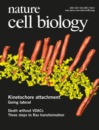
No. 5 May 2007
The kinetochore outer plate in PtK1 cells is a fibrous network (yellow) that forms multiple attachments to both the plus-end tips and the walls of spindle microtubules (red).
-
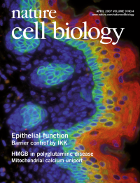
No. 4 April 2007
IKK1[letter p461]
-
No. 3 March 2007
[letter p310]
-
No. 2 February 2007
Chemotaxis in Dictyostelium is driven by the selective retention of randomly generated pseudopods.
-
No. 1 January 2007
Sharp Dpp boundaries sculpt fly legs by triggering cell death in the joints.