Volume 27
-
No. 12 December 2009
Artwork drawn by participants at an International Genetically Engineered Machine (iGEM ) competition, where undergraduates design, build and test simple biological systems made from standard, interchangeable biological parts (p.1099). This issue focuses on the emerging field of synthetic biology. Credits: Kim Caesar, based on a photograph provided by David Appleyard/iGEM.
Focus
-
No. 11 November 2009
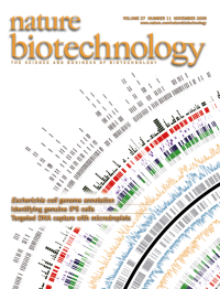
Graphical representation of part of the Escherichia coli genome. Cho et al. integrate several genomewide measurements of transcription and translation with the current annotation to elucidate the genomes architecture (p 1043). Credit: Byung- Kwan Cho/Kimberly Caesar.
-
No. 10 October 2009
Scanning electron micrograph of T lymphocytes attacking a cancer cell. Kortylewski et al. activate antitumor immunity using a CpG toll-like receptor 9 agonist linked to siRNA (p 925). Credit: Steve Gschmeissner, Science Photo Library.
-
No. 9 September 2009
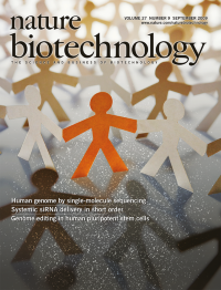
Fluorescently tagged nucleotides (small white 'dots') bound to complementary bases on immobilized single DNA molecules provide the backdrop for this image. With four week-long runs on one instrument, Pushkarev et al. sequence an individual's genome (p 847). Credits: Erin Dewalt, based on "One in a million" by Dimitri Vervitsiotis/Getty Images
-
No. 8 August 2009
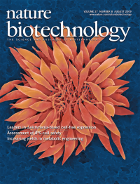
Scanning electron micrograph of Leishmania spp. Mureev et al. use species-independent translational leader sequences to develop a cell-free protein expression system based on Leishmania tarentolae cell extracts (p 747). Credit: Dennis Kunkel Microscopy, Inc.
-
No. 7 July 2009
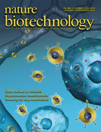
Sequential therapy of drug-resistant cancers with siRNA- and drug-containing minicells bearing O-polysaccharide chains (yellow). MacDiarmid et al. show that RNAi-mediated silencing of the gene encoding a multidrug resistance protein (magenta) reverses tumor drug resistance, increasing the efficacy of subsequent treatment with cytotoxic drugs (p 643). Credits: Martin Hale, © Animated Biomedical Productions, and Russell Kightley, © Russell Kightley Media.
-
No. 6 June 2009
Scanning electron micrograph of Pichia pastoris. De Schutter et al. present the complete genomic sequence of this important protein expression system (p 561). Credit: Dennis Kunkel Microscopy, Inc.
-
No. 5 May 2009
In silicopredicted behavior of four synthetic gene networks. Ellis et al. engineer networks with predictable behavior using libraries of experimentally and mathematically characterized promoters (p 465). Credit: Kimberly Caesar.
-
No. 4 April 2009
Padlock probes bound to bisulfitetreated genomic DNA in which cytosine residues (red) that are not methylated have been converted to uracils. Deng et al. and Ball et al. use customized padlock probes and next-generation sequencing to identify differences in DNA methylation between induced pluripotent stem cells and the fibroblasts from which they were derived (p 353, p 361).
-
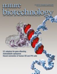
No. 3 March 2009
Artist's impression of a U1 adaptor oligonucleotide (orange) recruiting the U1 snRNP (snRNa in purple) to the 3' end of a target pre-mRNA (blue). Goraczniak et al. show that the resulting inhibition of transcript polyadenylation silences expression of the target gene (p 257). Credit: Ken Eward © Biografx, with data provided by Holger Stark.
-
No. 2 February 2009
Artist's rendering of streptavidincoated magnetic beads used to pull down ultra-long biotinylated RNA 'baits' designed to capture specific genomic DNA fragments. Gnirke et al. use the approach for targeted Illumina sequencing, represented by the processed image of a massively parallel sequencing experiment (p 182). Credit: Ken Eward ©BioGrafx.
-
No. 1 January 2009
A collage of seven images showing ~1.6 cm of the cervical and thoracic spinal cord of an adult mouse intravenously injected with AAV 9-GF P. Widespread transduction is seen in gray-matter astrocytes but not whitematter astrocytes (labeled with GFA P staining in blue) (p 59).