Volume 35
-
No. 12 December 2022
The cover shows GeoMx digital spatial profile analysis to identify tumor-infiltrating lymphocyte density-dependent immune-related gene expression signatures in microsatellite instability-high colorectal carcinomas. For more information see the paper by Jung et al, this issue, p 2018.
-
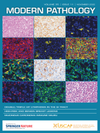
No. 11 November 2022
The cover shows double/triple hit lymphoma in the gastrointestinal tract. For more information see the paper by Guo et al, p 1667, this issue.
-
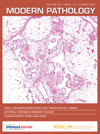
No. 10 October 2022
The cover shows hematoxylin and eosin well differentiated papillary mesothelial tumor for more information see the paper by Churg and Galateau-Salle, p 1327, this issue.
-
No. 9 September 2022
The cover shows unusual features of papillary renal neoplasm with reverse polarity, including cystic neoplasm filled with eosinophilic proteinaceous material containing loose hemosiderin-laden macrophages and cellular debris, edematous and expanded fibrovascular cores containing chronic inflammatory infiltrates, focal clear cell changes, and hobnail conformation. For more information, see the paper by Al-Obaidy et al, p. 1279, this issue.
-
No. 8 August 2022
The cover shows sample histologic images of training cohort tissue microarray spots containing cribriform glands and how cribriform size is measured. For more information, see the paper by Chan et al, p 1092, this issue.
-
No. 7 July 2022
The cover shows the histologic spectrum of endometrial carcinomas with HER2 amplification. For more information, see the paper by Ross et al, this issue (p 962).
-
No. 6 June 2022
The cover shows histological and Syn/periodic acid-Schiff double-staining of gastric amphicrine carcinoma. For more information, see the paper by Sun et al, this issue, p. 808.
-
No. 5 May 2022
The cover shows local extension of a testicular adult granulosa cell tumor. For more information see the paper by Siegmund et al, p 697, this issue.
-
No. 4 April 2022
The cover shows assessment of immune signatures in the tumor and tumor microenvironment of a chemotherapy-treated stage III colorectal cancer patient using an in situ multiplexed immunofluorescence imaging and single cell analysis technology (Cell DIVETM). For more information, see the paper by Stachtea et al,p 564, this issue.
-
No. 3 March 2022
The cover shows the main features of stroma of low-grade oncocytic renal tumor. For more information see the paper by Morini et al, p 352, this issue.
-
No. 2 February 2022
The cover shows Colocalization of osteopontin with known drusen components in human donor eyes. For more information, see the paper by Lekwuwa et al, p 165, this issue.
-
No. 1 January 2022
Cover: (Upper) DICER1 cystic hepatic neoplasm presenting as a cystic lesion. (Lower) Small intestinal polyp in a child with pleuropulmonary blastoma. For more information, see the paper by González et al, this issue, p 4.