
Figure 8 - Example of esophagopharyngeal regurgitation captured during prolonged manometric and dual pH recording.
From the following article
Clinical disorders of the upper esophageal sphincter
Ian J. Cook
GI Motility online (2006)
doi:10.1038/gimo37
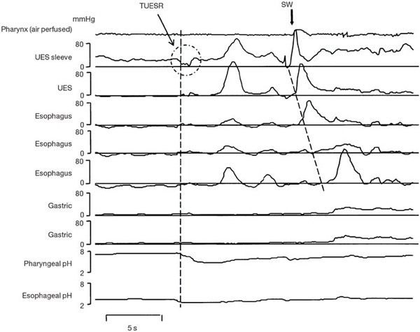
The sleeve sensor that was monitoring LES pressure has slipped into the stomach; hence, no recording of LES pressure is visible. However, during a period of prolonged esophageal acidification, this example clearly shows a spontaneous (non–swallow-related) UES relaxation, which mediates pharyngeal acidification.
Powerpoint slides for teaching
If the slide opens in your browser, Select "File > Save as" to save it.
Download Power Point slide (489K)