Figures, tables and video
From the following article
W.G. Paterson, Raj K. Goyal and Fortunée Irene Habib
GI Motility online (2006)
doi:10.1038/gimo20
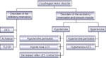
Figure 1
Pathophysiologic classification of motor disorders of smooth muscle portion of esophagus.
Full size figure and legend (37K)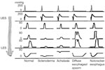
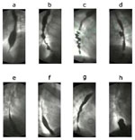
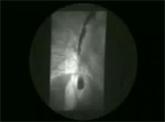
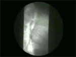
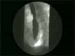
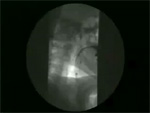
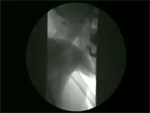
Figure 3
Radiologic appearance of some motor disorders of smooth muscle portion of the esophagus.
Full size figure and legend (22K)

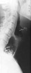

Figure 7
Illustration of hypotensive (incompetent) peristaltic contractions.
Full size figure and legend (49K)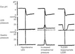
Figure 8
Three different mechanisms of LES incompetence in gastroesophageal reflux.
Full size figure and legend (38K)
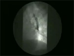
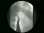
Figure 9
Scleroderma esophagus peptic stenosis as seen in a double-contrast examination of the esophagus.
Full size figure and legend (35K)
Table 1
Correlation of various parameters of esophageal motility disorders: major symptom, clinical syndrome, esophageal motility findings, esophageal bolus transport, pathophysiology or the anatomic site of major involvement
Full size table and legendTable 3
Summary of manometric findings in selected esophageal motor disorders
Full size table and legend