
Figure 3 - Radiologic appearance of some motor disorders of smooth muscle portion of the esophagus.
From the following article
W.G. Paterson, Raj K. Goyal and Fortunée Irene Habib
GI Motility online (2006)
doi:10.1038/gimo20
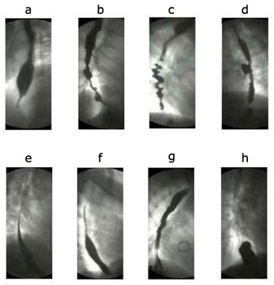
The figures are stills from the linked videos. a; Classical achalasia, showing a dilated esophageal body bird beak-like narrowing of lower esophageal sphincter (see Video 1). b; Vigorous achalasia showing diffuse spasm-like contractions in the esophageal body with closed lower esophageal sphincter (see Video 2). c; Diffuse esophageal spasm shows typical corkscrew appearance of the lower part of the esophagus (see Video 3). d: Midesophageal propulsion diverticulum with esophageal motility disorder (see Video 5). e; Normal peristaltic sequence for comparison with slow esophageal transit (see Video 6). f; Hypotensive (incompetent) esophageal peristalsis with slow esophageal transit through the esophagus (see Video 7). g; Gastroesophageal reflux (see Video 8). h; A sliding hiatal hernia (see Video 9). (Courtesy of Dr F.I. Habib)
Powerpoint slides for teaching
If the slide opens in your browser, Select "File > Save as" to save it.
Download Power Point slide (733K)