
Figures, tables and video
From the following article
Sphincter mechanisms at the lower end of the esophagus
Ravinder K. Mittal and Raj K. Goyal
GI Motility online (2006)
doi:10.1038/gimo14
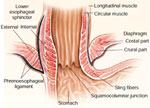
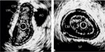
Figure 2
Ultrasonographic images of the esophagus (left) and lower esophageal sphincter (LES, right).
Full size figure and legend (50K)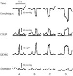
Figure 3
Esophagogastric junction pressure (EGJP) during diaphragmatic contraction recorded by a reverse perfused sleeve sensor equipped with electrodes to record electromyographic activity of the crural diaphragm.
Full size figure and legend (40K)
Figure 4
Reflex contraction of the esophagogastric junction recorded by a reverse perfused sleeve sensor equipped with electrodes to record crural DEMG activity.
Full size figure and legend (25K)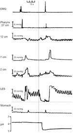
Figure 5
An example of swallow-induced lower esophageal sphincter (LES) relaxation (left) and transient LES relaxation (right).
Full size figure and legend (40K)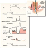
Figure 6
Physiologic record of a spontaneous, transient relaxation of the LES.
Full size figure and legend (64K)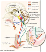

Table 1
Effects of some hormones and putative neurotransmitters on the lower esophageal sphincter and the possible sites of action
Full size table and legendTable 2
Pharmacologic agents known to inhibit transient lower esophageal sphincter relaxations
Full size table and legend