Thank you for visiting nature.com. You are using a browser version with limited support for CSS. To obtain
the best experience, we recommend you use a more up to date browser (or turn off compatibility mode in
Internet Explorer). In the meantime, to ensure continued support, we are displaying the site without styles
and JavaScript.
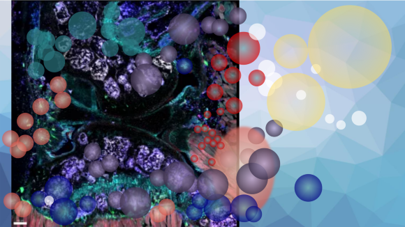
Visualising biology is a proven powerful strategy of scientific discovery, with live microscopy being a particularly successful endeavour. But only the latest revolutionary technologies in advanced live cell microscopy demonstrate the desired technological leap capable of overcoming the challenge to image the inner workings of living cells, tissues, and organs. The expectation is that these new technologies and methodologies will shape biomedical sciences in the years to come.
Communications Biology is inviting submissions on the topic of articles in live microscopy – from new tools to emerging techniques, from conventional to advanced light microscopy - with the aim of publishing high-quality research devoted to advance our understanding of biology. Reviews, Perspectives, and Comments covering these topics will also be considered for inclusion in the Collection. All submissions will be subject to the same review process and editorial standards as regular Communications Biology Articles.
This review introduces the basic principle of fluorescence and two-photon excitation microscopy that is aimed at novice and advanced microscopists highlighting current techniques, trends, and practical guides for live imaging optimization.
Communications Biology is inviting submissions on the topic of live microscopy – from new tools to emerging techniques, from conventional to advanced light microscopy - with the aim of publishing high-quality research devoted to advance our understanding of biology.
This Mini Review provides a focus on corneal confocal microscopy and insights into the neuroanatomy of the corneal surface using this technique and open debates are highlighted.
This Perspective looks at the choice of label-free optical microscopes available and discusses applications as well as scope for further improvement of labelfree imaging.
The no-reflow phenomenon after stroke is attributed to damaged spontaneous vasomotion of cerebral arterioles, which can be alleviated by maintaining mitochondria-ER contact-dependent Ca2+ oscillation.
The authors propose a new method for two-photon microscopy that can produce large cranial windows covering both the cerebrum and cerebellum of mice. The windows maintain transparency for over 5 months and allow multi-scale two-photon imaging of neuronal structures and functions in awake mice.
A high-resolution imaging apparatus that allows functional retinal imaging in awake and free moving mice was developed, allowing the study of the effect of anaesthesia on eye motion, blood flow and retinal thickness.
A live-cell trajectory embedding analysis characterizes the morphodynamical changes associated with molecular and ligand-induced responses and improves identification of metastable cell states compared to morphological snapshot-based analysis.
A workflow for 3D characterization of the mouse small intestine with optical projection tomography allows the identification of sparsely-distributed regions of interest in large volumes while retaining compatibility with high-resolution microscopy modalities.
An imaging platform with self-supervised deep learning allows for the imaging of cerebral blood flows under the effect of cocaine in awake mice using 3D ultrahigh-resolution optical coherence Doppler tomography.
A combination of live imaging and immunofluorescence on donor islet cells uncover an anti-correlation of enzyme-bound NAD(P)H and insulin secretion power.
Kinetics of cargo degradation in phagosomes are measured through engineered RotSensors, capturing their translational and rotational dynamics in parallel.
The physiological role of Vsp in zebrafish is assessed, revealing Vsp expression in the mid-intestine for dietary protein absorption. A comparative study on marine invertebrate Ciona intestinalis suggests conservation of Vsp function in the GI tract.
Hahn et al. present a method to visualize the endocrine human pancreas in 3D and calculate volumetric data. Using immunolabeling to visualize targets of interest and in reconstructing large tissue parts from imaged cm3-sized tissue blocks, they use their method to reveal previously unknown morphological differences in the endocrine pancreas affected with type 2 diabetes.
Allam, Hu, et al. present SpatialViz, a suite of algorithms to explore spatial relationships in multiplexed tissue images by visualizing and quantifying single-cell granularity and anatomical complexity in diverse multiplexed tissue imaging data. The authors employ SpatialViz on 20-plex protein data in tissue sections from normal and chronic tonsillitis cases and observe GrB and CD86 coexpression and CD3 + CD4+ enrichment in diseased tonsils compared to healthy tonsils, and demonstrate the utility of SpatialViz as a wide-application spatial visualization method.
Using phasor-FLIM (Fluorescence Lifetime Imaging Microscopy), Wang et al. reveal differential usages of oxidative phosphorylation and glycolysis in living islet cells following glucose stimulation. This study suggests the applicability of FLIM as a drug discovery tool as it reports perturbations in glucose metabolism in living islet cells.
Mehmet S. Ozturk et al. present an intravital mesoscopic fluorescence tomography (IFT) technique that is capable of tracking fluorescently labeled tumor cells inside living mice over several months. It demonstrates a promising new method for studying tumor growth dynamics in a quantitative and longitudinal fashion in-vivo.
This study presents an image processing-based stage height control and optical trap dithering technique to detect protein-protein interactions. It was used to show that the MT binding rate for KIF5B has a strong spatial and motor length dependence.
In this Perspective, the authors argue that it is time for biology to expand the traditional approach of observation with an engineering approach that transforms the nature of the underlying experiments to interactive context-depended design problems.
Content-aware image restoration (CARE) is extended to luminescence imaging allowing shorter exposure times thus improving temporal resolution and throughput.
A new instrument named OptoRheo combines light sheet fluorescence microscopy and particle tracking microrheology for live imaging and micromechanical sensing of extracellular matrix-cell interactions.
A high-throughput image-based approach quantifies single-cell biophysical fractal-related properties at subcellular resolution with statistical power for cell classification, drug response assays, and cell-cycle progression tracking.
Deep-Manager is a software platform that enables efficient selection of features with lower sensitivity to unspecified disturbances across sets of similar experiments in bioimaging.
A fast multidither coherent optical adaptive technology (fCOAT) focuses light through scattering tissue for optogenetic stimulation with subcellular resolution.
Raman spectroscopy is employed to show simultaneous, label-free imaging of RNA, DNA, and proteins, revealing secondary structural features of intracellular LLPS organelles, and condensed chromosome.
Bioluminescence microscopy with deep learning enables subsecond exposures for timelapse and volumetric imaging with denoising and yields high signal-to-noise ratio images of cells.
Using unsupervised image-to-image synthesis, homography regression-based co-registration of fluorescence-histology images is improved and can be used on various image formats across full-spectral emission wavelengths.
The development of an image splitter based multi-colour single-molecule localization microscopy method (splitSMLM) in combination with a spectral demixing algorithm improves localization accuracy as exemplified by three-colour imaging of nuclear pore complex proteins.
The high-speed single-molecule miRNA detection technique dynamic FRET-FISH shows increased detection speed and high target specificity, detecting a single miRNA in ten seconds.
A red fluorescent protein-based indicator of cyclic guanosine 3′, 5′-monophosphate (cGMP) allows for multi-colour imaging and optogenetic applications while monitoring cGMP activity in cells.
A deep learning approach, termed wrinkle force microscopy, allows for conducting traction force microscopy by observing bright-field cell images but without using its conventional requirements such as fluorescent microbeads and confocal microscopy.
A multiplexed machine-vision-based tracking system enables the high-content analysis of C. elegans behavior in several 96-well plates at a time and shows its utility for comparisons across multiple genotypes and upon drug treatment, tackling the bottlenecks in behavioural quantification.
Zhang, Redington & Gong develop FRET-based ratiometric calcium sensors with pairs of green and red fluorescent proteins. Their best performers, Twitch-GR and Twitch-NR inherit the superior photophysical properties of their constituent fluorescent proteins, enabling them to outperform existing ratiometric calcium sensors in brightness, photobleaching, and signal fidelity metrics.
Hedde et al. demonstrate the use of ultrafast phasor-based hyperspectral snapshot microscopy for biomedical imaging. This technique can improve imaging speed by 10-100 fold and enables 3D hyperspectral imaging of live tissues without using expensive and specialized hyperspectral cameras.