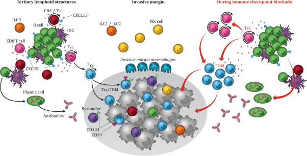
The schematic representation of the tumor immune environment shows the composition and function of a tertiary lymphoid structures (TLS), who are usually found peritumorally in the stroma and/or in the invasive margin. The chemokine CXCL13, produced by CD8+ T cells, induces chemotaxis by binding to the receptor CXCR5, mainly expressed by B cells and TFH cells, and regulates the organization of B cells inside the follicles of lymphoid tissues. The TLS consists out of a T cell-rich zone containing mature dendritic cells (DCs), in close proximity to GC containing follicle like-B cells, intermingled with follicular dendritic cells (FDCs) and surrounded by plasma cells and helper-innate lymphoid cell group 3 (ILC3) at the edge of the TLS. In the optimally organized TLS immune structure, DCs, FDCs, T cells and B cells interact and activate each other, promoting a local sustained immune response including the induction of T cell effector function, antibody generation, and clonal expansion. The stroma surrounding the tumor epithelium and the invasive margin further harbors cellular immune components including NK cells, macrophages, ILC1s and ILC2s, and a nonimmune cellular component, including fibroblasts. Within the tumor epithelium ILCs, NK cells, B cells, and different T cell subsets are present, including TEX cells,-tumor-specific CD103+CD39+ TRM CTLs and CD103+CD39- bystander TRM cells. Upon ICB, both T and B cell signaling increases. TCF1-expressing TPE cells expand and differentiate into TRM cells migrating to the tumor, where they can exert their cytolytic potential. The ICB response also increases B cell receptor diversity by means of SMH and CSR and induces their clonal expansion and differentiation into advanced antibody-producing plasma cells. TLS: tertiary lymphoid structure, TFH cells: follicular helper T cells, DCs: dendritic cells, GC: germinal center, FDCs: follicular dendritic cells, ILC3: helper-innate lymphoid cell group 3, NK cells: natural killer cells, ILC1: helper-innate lymphoid cell group 1, ILC2: helper-innate lymphoid cell group 2, TEX: terminally exhausted T cells, CTLs: cytotoxic lymphocytes, TRM: tissue resident memory, ICB: immune checkpoint blockade, TCF1: transcription factor 1, TPE: progenitor STEM-like exhausted cells, SMH: somatic hypermutation, RCS: recombinant class switch
- Sterre T. Paijens
- Annegé Vledder
- Hans W. Nijman