

© Mykyta Dolmatov/iStock/Getty Images Plus
A tooth requiring root canal treatment (RCT) has often previously experienced caries, trauma, or restoration. RCT further injures the tooth through multiple mediums including quantitative removal of tooth structure for endodontic access, qualitative deterioration of tooth structure via root canal irrigants, and de-innervation leading to reduced neurosensory feedback to occlusal loading. Subsequently, the root-filled tooth is at significantly increased risk of (non-restorable) fracture, and requires careful restorative consideration. Part 3 of this series outlines key factors to consider when planning restoration of root-filled posterior teeth.
-
1.
Saving or replacing an existing restoration
-
a.
A restorability assessment should be undertaken before RCT, and include removal of all existing restorative materials in order to be able to identify caries, cracks, and marginal leakage.1 However, exceptionally, when a tooth requires RCT treatment subsequent to recent placement of a restoration (within last six months), and the treating clinician is familiar with the amount, distribution, and quality of the tooth tissue that remained at the time of restoration, endodontic access can be prepared through an existing restoration. However, it should be borne in mind that creating an endodontic access cavity through an existing restoration (especially cast restorations) often hinders proper orientation, makes root canal identification more challenging, and increases the likelihood of excessive removal of dentine as well as the risk of perforation. Where a restoration has been compromised via preparation of an endodontic access cavity, it should be replaced following completion of the RCT.
-
b.
Where a tooth has an existing prosthetic crown, taking a pre-operative sectional silicone impression prior to the restorability assessment facilitates the subsequent manufacture of an indirect temporary restoration using a bis-acryl composite resin (eg ProTemp4, 3M ESPE, Seefeld, Germany).
-
a.
-
2.
Assessing tooth structure
-
a.
The margins of a cast restoration should ideally encircle the prepared tooth structure to create a ferrule. Desirable features of a ferrule have been reported to include: tooth tissue encircled by the ferrule should be >1 mm in height,2 >1.5 mm in thickness,3 and ideally extend circumferentially around the tooth.4 As a diagnostic aid, to determine whether an adequate ferrule effect can be generated, a crown margin may be prepared at the time of restorability assessment to quantify how much dentine remains.
-
b.
Where inadequate supra-gingival coronal dentine remains, additional height for the preparation may be gained via sub-gingival margin placement. The supracrestal attachment of the periodontium comprises both connective tissue and junctional epithelium, and has been reported to be 2.04 mm thick on average.5 Encroachment of the restorative margin into the supracrestal attachment is associated with inflammation +/- loss of periodontal supporting tissue.6 Therefore, the clinician should undertake 6-point probing around the tooth to establish the profile of the periodontal pockets and avoid placement of the margin closer than 0.5 mm from the depth of the gingival sulcus.7 Alternatively, surgical crown lengthening can be employed to increase the amount of available supra-gingival dentine available.
-
a.
-
3.
Timing
-
a.
It is the authors' opinion that the risk of fracture and possible subsequent need for extraction outweighs the risk of delaying definitive restoration to confirm apical healing. Indeed, it has been shown that delayed provision of a definitive restoration for root-filled teeth has been shown to be associated with increased risk of tooth loss.8 Therefore, where possible, a definitive core with cuspal-coverage should be placed at the obturation appointment, or as soon as possible after this. However, provision of a definitive cast restoration should be delayed if the tooth remains symptomatic (eg tenderness, non-healing sinus, or suppuration) following RCT, pointing to failure of the endodontic treatment, and need for root canal re-treatment +/- referral to a specialist endodontist.
-
b.
Where doubt exists as to whether the RCT has been successful, then a root-filled posterior tooth should be provisionally restored with either a definitive core incorporating cuspal-coverage, or a temporary crown, prior to definitive management.
-
a.
-
4.
Pulp chamber management
-
a.
Root filling material which extends into the pulp chamber should be removed prior to restoration of the tooth, in order to maximise the available space for retention of the core. Gutta percha (GP) can easily be easily removed with a heated plugger, or ultrasonic scaler (used for up to ten seconds at a time, without water coolant, in order to guard against the risk of thermal injury to the tooth and supporting tissues).
-
b.
Remnants of unset sealer on the floor and walls of the pulp chamber should be removed using a solvent such as pure eucalyptus oil, or an ethanol-based cleaner (eg AH Plus Cleaner, Dentsply, Konstanz, Germany) in conjunction with a cotton wool pledget held by tweezers, or a microbrush (eg Ultrabrush, Microbrush International, Grafton, USA).
-
c.
Where the root canals are not going to be used to contribute towards retention of the core (see next section), then root canal orifices should be sealed to reduce the risk of (re)contamination of root canal system in the event of future marginal breakdown around the core material. Rubber dam should remain in place until the orifices are sealed, and ideally until the completion of core placement. An orifice seal can be achieved by removing the coronal 2 mm of GP from the root canal orifice with a heated plugger, ultrasonic scaler, or (less ideally) a Gates Glidden drill. The coronal 2 mm of each root canal is then sealed with glass ionomer cement (GIC). Use of a resin-modified GIC provides the clinician with a level of control over the setting of the material, which can be initially manipulated with a Machtou plugger under illumination incorporating a blue-light filter; curing can be carefully accelerated by reflecting the unfiltered operating light into the pulp chamber (via a dental mirror) prior to further manipulation of the material, before commencing to final curing with a conventional light curing unit. Alternatively, if amalgam is to be used as the core material, then Intermediate Restorative Material (Dentsply Caulk, Milford, USA) can be placed which makes use of the inherent antibacterial properties of this zinc-oxide eugenol containing material.
-
a.
-
5.
Core placement
-
a.
Cavity preparation prior to core placement in a posterior tooth includes: removal of any unsupported enamel; use of the pulp chamber to enhance retention and resistance form; reduction of guiding cusps by 2 mm; reduction of balancing cusps by 1.5 mm; rounded internal angles; and, preparation of a 90° cavo-surface angle to create a 'butt finish'.
-
b.
Either amalgam or composite are advised as core materials in posterior teeth. Amalgam is a tried and tested material, offers good longevity, is strong in compression, offers good contrast with tooth tissue allowing ease of preparation, and is less technique-sensitive than composite alternatives. The mechanical retention of an amalgam core can be enhanced via extension of amalgam 2-4 mm into the orifice of each root canal. However, in a modified approach to that proposed by Nayyar,9 we advocate that to avoid further structural weakening of the tooth, root canals should not be further prepared to facilitate amalgam packing. Alternatively, retention may be enhanced via amalgam bonding.10
Where insufficient enamel remains for predictable enamel bonding, amalgam may be used to provide a definitive restoration
-
c.
Composite resin provides a contemporary alternative to amalgam. However, the challenges of achieving predictable light-activated polymerisation of resin monomers at depths greater than 5 mm have been reported.11 To benefit from the properties of a composite resin material, whilst negating the risk of incomplete polymerisation, the authors advocate the use of dual-cured composite resin for core build-up (eg ParaCore, Coltène/Whaledent AG, Altstätten, Switzerland). This material also has the following additional advantages: dentine bonding; efficient bulk-placement of material (as a monobloc), so saving time and increasing the likelihood that RCT and definitive core placement can be completed within a single-visit; can be prepared for an indirect restoration within four minutes of placement; and, the white colour contrasts with tooth tissue, which proves useful during tooth preparation procedures.
-
a.
-
6.
Definitive direct vs indirect restorations
-
a.
Cast restorations are recommended for the definitive restoration of root-filled molar teeth12,13 (Figures 1, 2, 3 and 4 show some of the stages in restoration of a root-filled 37 tooth). However, compared to molars, premolars have relatively thinner axial walls. Therefore, preparation of a premolar for a given type of indirect restoration will result in a proportionally greater loss of coronal tooth tissue compared to the same type of preparation in a molar tooth. Where insufficient coronal tissue remains to retain a core in a premolar, a post will often be required.
Fig. 1 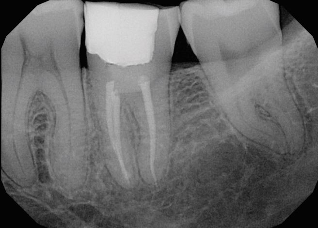
Post-operative long cone periapical radiograph of a 37 tooth following completion of root canal treatment and placement of a composite resin core
Fig. 2 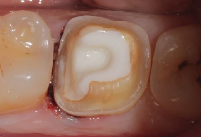
Crown preparation 37 tooth. Box cut within the confines of the composite resin core to enhance retention and resistance form
Fig. 3 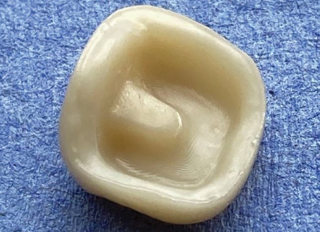
Full-coverage, monolithic, zirconia crown for 37 tooth (detail of fitting surface)
Fig. 4 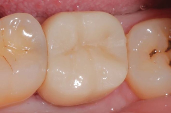
Fitted zirconia crown
-
b.
As an alternative to a cast restoration, where a circumferential ring of enamel remains, a definitive cuspal-coverage direct composite resin restoration may be placed. Alternatively, where insufficient enamel remains for predictable enamel bonding, amalgam may be used to provide a definitive cuspal-coverage restoration.
-
a.


-
7.
Use of posts in posterior teeth
-
a.
Post placement to retain cores in molar teeth is not necessary,14 and should be avoided, as it can weaken teeth, predispose to root fracture, or result in perforation. Therefore, methods to enhance retention of the core in a molar should be limited to a modified Nayyar core, or use of a bonded amalgam or composite material, as previously outlined.
-
b.
In contrast, fibre posts in root-filled premolar teeth have been reported to be beneficial in aiding core retention where two or fewer axial walls remain.15 However, there is a negative correlation between the amount coronal tooth structure remaining, and the likelihood of mechanical failure of a fibre post treated tooth.16 The inherent flexure of fibre posts can precipitate marginal leakage, caries, and ultimately fracture of the core. Where a ferrule is not achievable, or as an alternate to fibre posts, clinicians may opt to place a cast precious metal post and core prior to crowning the tooth.17 The criteria for assessment and placement of posts was discussed in Part 2 of this series.
-
a.
-
8.
Design considerations for indirect restorations
-
a.
The preparation of posterior root-filled teeth for indirect restorations should be conservative, whilst meeting the requirements for cuspal-coverage, and adequate retention/resistance form. For example, an onlay design should be chosen where the buccal and lingual/palatal walls are intact. However, where three walls are compromised, but one proximal wall remains, a three-quarter crown may be appropriate. Only where all four axial walls have been previously compromised is a full-veneer preparation recommended.
-
b.
Where a root-filled posterior tooth with a Class I access cavity has a circumferential band of enamel remaining, an 'endocrown' can provide a very conservative cast cuspal-coverage solution.18 Endocrowns are monolithic porcelain (or gold) cast cuspal-coverage restorations, which utilise the pulp chamber for retention, and have been reported to provide a high-level of success at ten years.19
-
a.
-
9.
Material choice for indirect restorations
-
a.
Gold alloys (type III or IV) are excellent for cuspal-protection of root-filled teeth, owing to their strength in thin section (facilitating conservative tooth preparations), and capacity for bonding to tooth tissue. However, patients may not accept gold in the aesthetic zone.
-
b.
Lithium di-silicate glass ceramic restorations (eg eMax, Ivoclar Vivadent, Schaan, Lichtenstein) are not as strong as gold, but have excellent aesthetic properties and can be bonded to tooth tissue, so facilitating a conservative tooth preparation.
-
c.
Monolithic yttrium-stabilised zirconia restorations (eg Procera, Nobel Biocare, Kloten, Switzerland) are stronger than lithium disilicate restorations, but when finished with an overlying layer of feldspathic porcelain, the fracture resistance of the restoration drops to that of the weaker material (eg the feldspathic porcelain). Zirconia cannot be predictably bonded to tooth tissue, and so carries the disadvantage of requiring a conventional tooth preparation to derive the necessary retention and resistance form. Adjustment of the crown creates a roughened surface which if not adequately polished can abrade the opposing tooth. If the patient is a bruxist and will not tolerate gold, monolithic zirconia would be an acceptable choice for a crown on a posterior tooth.
-
a.
As stated previously, these top tips are based mainly from experience and may need some adaptation to an individual's circumstances and practice. We hope this series will be a useful reference for managing restoration of root-filled teeth.
References
Abbot P. Assessing restored teeth with pulp and periapical diseases for the presence of cracks, caries and marginal breakdown. Aust Dent J 2004; 49: 33-39.
Sorensen J, Engleman M. Ferrule design and fracture resistance of endodontically treated teeth. J Prosthet Dent 1990; 63: 529-536.
Haralur A, Al-Qahtani A, Al-Qarni M, Al-Homrany R, Aboalkhair A. Influence of remaining dentin wall thickness on the fracture strength of endodontically treated teeth. J Conserv Dent 2016; 19: 63-67.
Arunpraditkul S, Saengsanson S, Pakviwat W. Fracture resistance of endodontically treated teeth: three walls versus four walls of remaining coronal structure. J Prosthodont 2009; 18: 49-53.
Gargiulo A, Wentz F, Orban B. Dimensions and relations of the dentogingival junction in humans. J Periodontol 1961; 32: 261-267
Jepsen S, Caton J, Albandar J et al. Periodontal manifestations of systemic diseases and developmental and acquired conditions: Consensus report of workgroup 3 of the 2017 World Workshop on the Classification of Periodontal and Peri-Implant Diseases and Conditions. J Clin Periodontol 2018; 45: 219-229.
Kois J. The restorative-periodontal interface: biological parameters. Periodontol 2000 1996; 11: 29-38.
Lynch C, Burke F, Ríordáin R, Hannigan A. The influence of coronal restoration type on the survival of endodontically treated teeth. Eur J Prosthodont Restor Dent 2004; 12: 171-176.
Nayyar A, Walton R, Leonard L. An amalgam coronal-radicular dowel and core technique for endodontically treated posterior teeth. J Prosthet Dent 1980; 43: 511-515.
Sagsen B, Aslan B. Effect of bonded restorations on the fracture resistance of root filled teeth. Int Endod J 2006; 39: 900-904.
Herrero A, Yaman P, Dennison J. Polymerization shrinkage and depth of cure of packable composites. Quintessence Int 2005; 36: 25-31.
Aquilino S, Caplan D. Relationship between crown placement and the survival of endodontically treated teeth. J Prosthet Dent 2002; 87: 256-263.
Ng Y-L, Mann V, Gulabivala K. A prospective study of the factors affecting outcomes of non-surgical root canal treatment: part 2: tooth survival. Int Endod J 2011 44: 610-625.
Sorensen J, Martinoff J. Intracoronal reinforcement and coronal coverage: a study of endodontically treated teeth. J Prosthet Dent 1984; 51: 780-784.
Ferrari M, Vichi A, Fadda G et al. A randomized controlled trial of endodontically treated and restored premolars. J Dent Res 2012; 9: 72-78.
Ferrari M, Cagidiaco M, Goracci C et al. Long-term retrospective study of the clinical performance of fibre posts. Am J Dent 2007; 20: 287-291.
Sarkis-Onofre R, Fergusson D, Cenci M, Moher D, Pereira-Cenci T. Performance of post-retained single crowns: a systematic review of related risk factors. J Endod 2007; 43: 175-183.
Bindl A, Mörmann W. Clinical evaluation of adhesively placed Cerac endo-crowns after 2 years - preliminary results. J Adhes Dent 1999; 1: 255-265.
Belleflamme M, Geerts S, Louwette M, Grenade C, Vanheusden A, Mainjot A. No post - no core approach to restore severely damaged posterior teeth: An up to 10-year retrospective study of documented endocrown cases. J Dent 2017; doi: 10.1016/j.jdent.2017.04.009.
Author information
Authors and Affiliations
Rights and permissions
About this article

Cite this article
Smith, R., McColl, E. & Bryce, G. Top tips for restoration of root-filled teeth: Part 3: posterior teeth. Br Dent J 233, 830–832 (2022). https://doi.org/10.1038/s41415-022-5292-5
Published:
Issue Date:
DOI: https://doi.org/10.1038/s41415-022-5292-5
This article is cited by
-
Top tips for endodontic access
British Dental Journal (2024)
