
Abstract
Social behaviors, how individuals act cooperatively and competitively with conspecifics, are widely seen across species. Rodents display various social behaviors, and many different behavioral paradigms have been used for investigating their neural circuit bases. Social behavior is highly vulnerable to brain network dysfunction caused by neurological and neuropsychiatric conditions such as autism spectrum disorders (ASDs). Studying mouse models of ASD provides a promising avenue toward elucidating mechanisms of abnormal social behavior and potential therapeutic targets for treatment. In this review, we outline recent progress and key findings on neural circuit mechanisms underlying social behavior, with particular emphasis on rodent studies that monitor and manipulate the activity of specific circuits using modern systems neuroscience approaches. Social behavior is mediated by a distributed brain-wide network among major cortical (e.g., medial prefrontal cortex (mPFC), anterior cingulate cortex, and insular cortex (IC)) and subcortical (e.g., nucleus accumbens, basolateral amygdala (BLA), and ventral tegmental area) structures, influenced by multiple neuromodulatory systems (e.g., oxytocin, dopamine, and serotonin). We particularly draw special attention to IC as a unique cortical area that mediates multisensory integration, encoding of ongoing social interaction, social decision-making, emotion, and empathy. Additionally, a synthesis of studies investigating ASD mouse models demonstrates that dysfunctions in mPFC-BLA circuitry and neuromodulation are prominent. Pharmacological rescues by local or systemic (e.g., oral) administration of various drugs have provided valuable clues for developing new therapeutic agents for ASD. Future efforts and technological advances will push forward the next frontiers in this field, such as the elucidation of brain-wide network activity and inter-brain neural dynamics during real and virtual social interactions, and the establishment of circuit-based therapy for disorders affecting social functions.
Introduction
Individuals of certain organism species act cooperatively and competitively with peers for reproduction and survival. The ubiquity of such social behavior across various species, ranging primarily from insects to humans, suggests that it is evolutionarily advantageous. Social behavior includes various communications and interactions between two or more individuals of the same species [1]. When two conspecific individuals encounter each other, their interaction starts with an appetitive phase that involves detecting, approaching, and investigating the target individual as a social stimulus, followed by a consummatory phase that consists of stereotyped motor patterns that give rise to goal-directed actions such as aggression, mating, or parenting (Fig. 1A) [2, 3]. Although an aspect of social behavior is considered fundamentally innate and stereotypical, it is modulated by learning and memory to support its flexibility. Social behavior can also be categorized as affiliative or aggressive from the perspective of whether it has a positive and friendly nature or an intention to cause harm [4].

A A simplified stream of mouse social interaction. B Social preference and social recognition. See main text for details. The figure was created using Biorender.com.
Social behavior is mediated by a distributed large-scale network of multiple brain structures. Expression of social behaviors is highly susceptible to dysfunction of this network, which is commonly observed in individuals with abnormal neurological and neuropsychiatric conditions such as stroke, schizophrenia, and autism spectrum disorder (ASD) [5,6,7]. ASDs are common early-onset neuropsychiatric conditions characterized by social communication deficits and restricted and repetitive patterns of sensory-motor behaviors. Although many non-genetic factors have been linked with ASDs, decades of research have established a strong causative relationship with genetics. Supporting its symptomatic heterogeneity manifesting as a broad spectrum, multiple types of genetic abnormalities have been associated with ASDs [8]. These include a large number of single genes, a major subset of which encodes synaptic molecules. In addition, multiple genetic abnormalities, including various copy number variants (CNVs) produced by deletion or duplication of chromosomal fragments [9], are heavily implicated in the pathogenesis of ASD. Due to the complex genetic nature of the disorder and the difficulty associated with studying how each (or groups of) genetic abnormalities contribute to ASD symptomology, investigating mouse models that mimic the genetic and clinical features of ASD thus provides a promising avenue toward elucidating mechanisms of abnormal social behavior and potential therapeutic targets for treating this disorder.
Studies have used many different behavioral paradigms to establish the social tendencies of rodents and the underlying neural circuit bases. For example, in an appetitive phase of social interaction, rodents show an inherent sociable tendency to prefer investigating a conspecific rather than an inanimate object, termed social preference (Fig. 1B). Rodents can also discriminate against each social target since they spend more time exploring novel individuals than familiar ones—an ability referred to as social recognition or social memory. Thus, investigation time decreases as they explore the same social target multiple times. Rodents also exhibit behavioral manifestations of empathy, such as observational pain, and prosocial behavior, such as consolation of stressed conspecifics [4, 10].
Here, we review recent progress and findings on neural circuit mechanisms underlying social behavior and circuit defects responsible for impaired social behavior in various ASD models, particularly with emphasis on rodent studies that use systems neuroscience approaches at the circuit level. Since excellent reviews on consummatory behaviors and their underlying mechanisms are already available [1,2,3], we primarily focus on the appetitive phase of social interaction in this review. We overview a set of brain networks that mediate social behaviors and their dysfunction in ASD model mice, specifically the neural circuits involving the medial prefrontal cortex (mPFC), the anterior cingulate cortex (ACC), the insular cortex (IC), and the neuromodulatory systems. Although an exhaustive listing of all relevant literature exceeds the scope of this review, we summarize some remarkable findings obtained from the recent studies of social behavior deficits of ASD mouse models in Table 1. Finally, we propose some major outstanding questions for future research in this field.
Cortical control of social behavior
mPFC
Function in social behavior
Neurons in the mPFC form reciprocal networks with various subcortical regions such as the nucleus accumbens (NAc), amygdala, ventral tegmental area (VTA), and raphe nucleus [11], and crucially regulates cognition, emotion, and behavior [12, 13]. Although the involvement of the mPFC in social behaviors is well-established [14], the functional significance of the overall excitability of mPFC neurons in social behavior is not straightforward to understand. While the firing rate of some mPFC neurons increases when a mouse approaches a stranger [15], optogenetically-induced sustained elevation of mPFC pyramidal neuron excitability reduces social interaction behavior [16]. Consistently, distinct neuronal ensembles that are activated (ON ensembles) or suppressed (OFF ensembles) during social interaction carry information on social salience and novelty in the mPFC [17], suggesting that mPFC neurons that are implicated in social behavior are composed of heterogeneous populations.
Accumulating evidence indicates that mPFC controls social behaviors in a subregion- and projection target-specific manner (Fig. 2). Activation of projection terminals of the prelimbic (PL; an mPFC subregion) neurons at the NAc suppresses social preference [18]. During social interaction, the activity of basolateral amygdala (BLA)-projecting infralimbic (IL; another mPFC subregion) neurons is more prominent than BLA-projecting PL neurons, and inhibition of the IL-BLA pathway or activation of PL-BLA pathway reduces social behavior [19]. By contrast, a study that examined the role of opposite projections from the BLA to mPFC (including both PL and IL) demonstrated that activation of this pathway suppressed social interaction, whereas inhibition facilitated it [20], although this study did not discriminate between the BLA projections to the PL and IL. Another study showed that, during social interactions with a target mouse, dorsal mPFC–BLA coherence of local field potentials at the 4–7 Hz band is higher during leaving behavior compared to approach behavior [21], suggesting that rapid functional connectivity changes between the two areas accompany distinct aspects of social interaction.

A Representative cortical and subcortical regions and their projections that promote social interaction (blue) and those that regulate pain and fear transfer (black dash). B Representative cortical and subcortical regions and their projections that suppress social interaction (red). See main text for details. ACC anterior cingulate cortex, AMY amygdala, DRN dorsal raphe nucleus, IC insular cortex, IL infralimbic area, LHb lateral habenula, mPFC medial prefrontal cortex, NAc nucleus accumbens, PL prelimbic area, pPVT posterior paraventricular thalamus, PVN paraventricular nucleus of hypothalamus, VTA ventral tegmental area.
The local circuit mechanisms regulated by the inhibitory interneurons also contribute to the intricate control of social behavior. Parvalbumin (PV)-positive interneurons target their axons to pyramidal neurons’ soma and suppress their excitability. Activation of PV-positive interneurons partially rescues a disruption in social preference elicited by elevated excitability of mPFC pyramidal neurons via normalizing the excitatory/inhibitory (E/I) balance [16]. Furthermore, social interaction increases low gamma oscillations and firing rates of PV-positive but not somatostatin (SST)-positive interneurons in the mPFC [22]. Conversely, inhibition of PV-positive but not SST-positive interneurons reduces low gamma power and impairs social interaction [22]. Surprisingly, however, activation of either PV-positive or SST-positive interneurons at low gamma frequency produces a prosocial effect [22], demonstrating the difference and commonality of two distinct classes of interneurons in local circuit control of social behavior.
Encounters with same-sex and opposite-sex conspecifics activate shared and distinct brain-wide networks [23], which can lead to different consummatory social behaviors such as fighting, mating, and parenting, according to the sex of the two individuals [1,2,3]. Sex-specific circuits and their activity thus mediate aspects of social behavior, and consequently, circuit dysfunction in ASD may also reflect such male-female circuit-level differences. In the mPFC of female mice, silencing a subpopulation of SST-positive interneurons that express oxytocin receptors (OTRs) resulted in the loss of social interest in male mice, specifically during estrus [24], demonstrating an example of local circuit control of sex-specific social behavior.
The mPFC also mediates adverse effects of social isolation stress. The activity of mPFC neurons that project to the posterior paraventricular thalamus (pPVT) is enhanced during social interaction, and suppression of this projection reduces sociability [25]. Interestingly, juvenile social isolation stress, known to induce sociability deficits in adulthood, impairs the activation of this pathway upon social exposure. Moreover, activating this pathway in adulthood rescues sociability deficits caused by juvenile isolation. Early social isolation also impairs social recognition through influence on the excitability of IL but not PL neurons [26]. Specifically, mice that experienced early social isolation lack the increased activity of NAc-projecting IL neurons during interaction with familiar conspecifics. Furthermore, inhibition of this pathway impairs social recognition without affecting social preference, and likewise, activation of this pathway rescues social recognition deficit in socially isolated mice [26]. Besides these circuits, recent whole-brain screening using rodent functional magnetic resonance imaging (fMRI) combined with chemogenetics identified lateral habenula (LHb) as a brain region that responds to PFC hyperactivity [27]. The role of LHb in regulating social behavior is demonstrated by the evidence that the activation of LHb neurons or PFC terminals in the LHb suppresses social preference. Since LHb is known to be involved in stress response and depression [28], elucidating whether this circuit also controls response to social stress is an important future question.
Despite the accepted importance of mPFC in controlling social behavior, many findings suggest that its distinct neural pathways are engaged in various contexts of social behavior. Thus, deciphering how the entire mPFC circuits operate during social interaction is still underway. A particular difficulty with this line of investigation arises from the complexity that mPFC also serves various non-social functions such as processing internal and external cues, decision-making, and flexible goal-directed behavior [14, 29]. The functional role of mPFC in the social domain is likely mediated by these general cognitive functions common to the non-social domain.
Implication in ASD model mice
As we saw earlier, the mPFC-BLA circuit is one of the important pathways for the control of social behavior [19, 30]. Several ASD mouse models exhibit abnormal social behavior and deficits in this circuit, suggesting that functional alterations in this pathway are crucially involved in social dysfunctions in ASD. SHANK genes encode a family of postsynaptic scaffold proteins associated with ASD [31]. Selective genetic deletion of Shank3 in BLA-projecting mPFC neurons leads to elevated neural activity of this pathway and disrupts sociability [32]. The authors further show that similar PFC-BLA functional hyperconnectivity is also observed in individuals clinically diagnosed with ASD [32].
Maternal immune activation (MIA) and postnatal immune activation (PIA) by prenatal polyinosinic: polycytidylic acid-induced neuroinflammation in pregnant mice and early postnatal lipopolysaccharide injections, respectively, both result in social dysfunction and altered functions of mPFC-BLA pathway [33]. However, these two models have different cellular targets within this circuit. While MIA increases synaptic strength in glutamatergic mPFC projections to the BLA, PIA decreases feedforward GABAergic inhibitory postsynaptic responses in local BLA circuitry [33].
PTEN encodes a phosphatase that negatively regulates the PI3K-Akt-mTOR pathway, and germ-line heterozygous mutations of this gene are identified in individuals with ASD and macrocephaly [34]. PTEN heterozygous knockout mice display hyperconnectivity of mPFC to BLA projections, enhanced activity of mPFC and BLA in response to social stimuli, and social behavioral impairments [35]. The functional and behavioral abnormalities are reversed by pharmacological inhibition of S6 kinase beta-1 during development or by reducing the activity of the mPFC–BLA circuit in adulthood [35].
Neurofibromatosis type 1 (NF1) is a disorder that has a high prevalence of ASD, and mice lacking a single NF1 allele show deficits in long-term social learning and increased activation of mitogen-activated protein (MAP) kinase pathway in neurons from BLA and PFC [36]. These mice also display elevated GABA and glutamate neurotransmission and long-term potentiation in the BLA, and the social learning deficits are rescued by pharmacological blockade of p21 protein-activated kinase in the BLA [36].
Interplays between genetic and epigenetic mechanisms play an essential role in social functions and their deficits in ASD model mice [37]. A study has shown that Shank3 deficiency induces the histone deacetylase HDAC2 upregulation via a β-catenin–dependent mechanism, and its knockdown in the mPFC or treatment with the HDAC inhibitor romidepsin rescues the social deficits of heterozygous Shank3-deficient mice [38]. These findings underscore the likelihood of an epigenetic mechanism underlying social defects associated with Shank3 deficiency.
ACC
Function in social behavior
The ACC in humans and rodents are highly interconnected with various brain areas primarily involved in emotional information processing, motivation, and autonomic function, such as orbitofrontal cortex, amygdala, NAc, hypothalamus, and autonomic brain stem nuclei [39]. The ACC has been implicated in various aspects of social cognition [40] – its activity is associated with other-referenced rewards [41] and negative valuations such as social anxiety [42, 43]. The ACC is also involved in the processing of the affective state of others [44]. Watching and learning are strategies to recognize cues for reward or punishment from others [45]. When observing another demonstrator mouse receiving an electric footshock, the observer mice show freezing behavior without experiencing the footshock, a process called observational fear learning [46]. Inactivation of ACC and an ACC-limited deletion of Cav1.2 calcium channel impaired this observational fear learning [47]. The ACC mediates these various types of social cognition processes via distinct projections. Recent studies demonstrate that the ACC-BLA circuit plays a crucial role in routing socially acquired aversive cue information and acquisition of observational fear conditioning [48] and that hippocampus-dependent 5–7 Hz oscillations in the ACC-BLA circuit in the right hemisphere are essential in empathic fear [49]. Furthermore, the ACC-NAc circuit regulates the social transfer of pain and analgesia, while the social transfer of fear depends on ACC-BLA circuit [50].
Implication in ASD model mice
Few studies have so far investigated the role of ACC in ASD model mice. Recently, a study has revealed morphological changes, hypoactivity, and weakened synaptic functions of pyramidal neurons in the ACC of mice deficient in Shank3 [51], revealing this region’s possible implications in ASD neurobiology. The study further demonstrated that selective deletion of Shank3 in the ACC resulted in synaptic dysfunction and social interaction deficits [51]. Moreover, optogenetic activation of ACC neurons, adult re-expression of Shank3, or pharmacological enhancement of α-amino-3-hydroxy-5-methyl-4-isoxazole propionic acid (AMPA)-type glutamate receptors improved social behavior in Shank3 mutant mice [51]. Interestingly, activation of ACC pyramidal neurons in Shank3 mutant mice rescued anxiety-like behavior. In contrast, their inhibition in wild-type mice did not affect it, indicating a possible dissociation of anxiety-like behavior and social dysfunction despite a proposed link between them [51].
IC
Basic anatomy and function
In this review, we draw particular attention to the IC as a unique cortical area that is still relatively understudied in the context of its involvement in the social behavior of rodents. The IC lies deep hidden below the frontal, parietal, and temporal lobes within the lateral sulcus in humans, and it is exposed mostly dorsal to the rhinal fissure and crossed by the middle cerebral artery on the lateral surface of the neocortex in rodents [52, 53]. Across species, the IC is subdivided along the rostrocaudal axis into two parts: the anterior insula and the posterior insula. In rodents, the IC is organized into three subdivisions arranged from dorsal to ventral and progressively devoid of the layer 4 granular layer: the granular subdivision, the dysgranular subdivision, and the agranular subdivision (Fig. 3A) [52, 54]. The IC forms an anatomic center with reciprocal connections to sensory, emotional, motivational, and cognitive systems, including the sensory and frontal cortices, amygdala, thalamus, and NAc, as well as with neuromodulatory inputs [55]. Rodent studies revealed that IC is involved in a wide variety of functions, including multisensory integration [56, 57], interoception [58, 59], pain [60, 61], taste [62,63,64], memory [65,66,67], emotion [58, 68], motivation [69], valence coding [70], physiological needs such as thirst and hunger [71, 72], aversive state processing [73, 74], and social functions such as social interaction [75, 76] and empathy [77,78,79]. In humans, the insula is also involved in self-awareness [80] and constitutes a part of the salient network that acts to detect novel and behaviorally relevant stimuli [81]. Atypical activation and connectivity of the IC are linked with various neuropsychiatric disorders such as schizophrenia and ASD [82, 83].

A Anatomical organization of mouse IC. AI agranular IC, DI dysgranular IC, GI granular IC. B IC as a link between sensory inputs and social decision-making network. AH anterior hypothalamus, BLA basolateral amygdala, BNST bed nucleus stria terminalis, HIP hippocampus, LS lateral septum, MeA medial amygdala, mPOA medial preoptic area, NAc nucleus accumbens, PAG periaqueductal gray, Str striatum, VMH ventromedial hypothalamus, VP ventral pallidum, VTA ventral tegmental area. C Microendoscopic calcium imaging from the agranular IC (AI) in freely-moving mice. The activity of neurons labeled with the green fluorescent calcium indicator protein GCaMP6f is imaged using a miniaturized head-mounted fluorescence microscope through a chronically implanted GRIN lens. D Social interaction in a home cage. A male subject mouse with a microscope attached to its head is allowed to interact with a male conspecific stranger mouse in its home cage. E Example GCaMP fluorescence traces of social-ON cells and a social-OFF cell imaged during a home cage session. The periods of social interaction are indicated in green. F Anatomical distribution of social-ON cells (red) and social-OFF cells (blue) in a microendoscopic field of view. C–F are adapted from [75].
The anterior and posterior insulae are closely related by intensive intra-insular connectivity but differ in connectivity to other brain regions [84]. The posterior insula is thought to be a major site for receiving and processing visceral, gustatory, and other bodily and sensory signals and affective states, whereas the anterior insula may serve more like a higher association cortex that integrates the information from the posterior insula with top-down and valence signals to guide motivated behavior through its downstream targets such as the ventral striatum and the motor cortex [84].
Social behavior requires multiple steps of information processing where multimodal sensory signals are integrated with interoceptive and affective information to select an adaptive behavioral response. The IC is anatomically situated to influence the activity of a large-scale network called the social decision-making network (SDMN). The SDMN is proposed to be an evolutionarily conserved network of brain structures that regulates adaptive social behavior in response to salient environmental stimuli [85, 86]. The SDMN consists of the core nodes of the social behavior network (SBN), which includes lateral septum (LS), medial preoptic area, anterior hypothalamus, ventromedial hypothalamus, periaqueductal grey, medial amygdala (MeA), and bed nucleus stria terminalis (BNST) [87], and the mesolimbic reward system that includes NAc, VTA, ventral pallidum, BLA, hippocampus, LS, BNST, MeA, the last three of which are overlapping nodes with the SBN (Fig. 3B) [85, 88]. Each of the SBN nodes is reciprocally connected with all the others, is sensitive to gonadal steroids, and participates in more than one social behavior [87]. The mesolimbic reward system evaluates stimulus salience via dopaminergic signaling and thus mediates rewarding aspects of social interactions [85]. The SDMN can thus assess and respond to a social environment with adaptive behavioral decisions by interacting with these brain regions. The IC receives sensory inputs via direct thalamic and cortical afferents and is connected bidirectionally or unidirectionally with most nodes of the SDMN [84, 88]. Consistently, a network analysis of immediate early gene expression after social interactions with conspecifics demonstrates that the IC acts at the interface of “social” and “emotional” modules of the SDMN in the rat [77].
Function in social behavior
In line with the hypothesis that the IC links multimodal sensory processing and social decision-making, accumulating evidence broadly supports the idea that the IC is causally implicated in regulating social behavior. Pharmacological blockade of NR2B subunit-containing N-methyl-D-aspartate (NMDA)-type glutamate receptors in the agranular IC (aIC) decreased social investigation of male rats [89]. Activation of the pathways from posterior IC to central nucleus of the amygdala but not NAc interrupts ongoing social interaction, potentially due to behavioral inhibition by the anxiety signals mediated by this pathway [73]. Social stimuli activate vasoactive intestinal peptide (VIP)-expressing interneurons in anterior IC, and inhibition of this neuron subtype impairs social preference [76]. Moreover, a recent study uncovered that the IC directly participates in the coding of social exploration behavior [75]. In this study, microendoscopic calcium imaging visualized the activity of aIC neurons while the subject mouse interacted with a novel conspecific mouse in the home cage (Fig. 3C, D). This study identified two groups of aIC neurons that exhibited social interaction-related activity—a larger fraction of cells (social-ON cells) that were more active and a smaller fraction of cells (social-OFF cells) that were less active during social exploration (Fig. 3E, F). Interestingly, social-ON cells responded to mice regardless of their positions and consisted of multiple subsets of cells, each of which was preferentially active during exploration under a particular behavioral state (i.e., moving or stationary) or with a particular target of physical contact (i.e., nose, body, or anus). These findings suggest that neuronal ensembles in the aIC encode the ongoing status of social exploration at an individual cell level and the social salience of the interaction target at a population level. Elucidation of the projection targets and the function of insular social-ON and social-OFF cells is of particular interest and awaits future investigations.
The IC also plays a critical role in empathy. Empathy is the ability to perceive others’ emotional states and to understand others’ points of view [44]. It is common across many species, such as humans, apes, elephants, dolphins, and rodents [44], and is thought to be a major motivational drive for affiliative prosocial behaviors that benefit other individuals [4]. Empathy is impaired in various psychiatric, neurological, and neurodevelopmental conditions, including psychopathy and ASD [90]. While humans likely exhibit the most complex forms of prosocial behaviors, rodents also display a variety of behavioral manifestations of empathy, such as social modulation of pain sensitivity in mice [79, 91, 92], observational fear responses to footshocks to another mouse [47], allogrooming toward stressed conspecifics in prairie voles and mice [93,94,95] and helping of trapped conspecifics in rats [96, 97].
Recent studies have identified several key brain areas implicated in empathy [4, 44, 98]. The anterior IC, in conjunction with the ACC (see also above section on ACC), forms the most critical network nodes for both affective and cognitive aspects of empathy. In humans, the anterior IC is activated by both directly experienced and empathic pain [98, 99] and by feeling and observing disgust [100]. Patients with lesions in the anterior IC display deficits in empathic pain perception [101]. Moreover, individuals with ASD show behavioral deficits in inferring others’ social emotions and reduced activities in the right anterior IC [102]. In rodents, allogrooming towards stressed conspecifics increases c-Fos expression in OTR-expressing neurons in the IC in mice [94], and inactivation of the IC prevents socially-elicited hyperalgesia in mice [92]. Glutamatergic projection from the IC to BLA and synaptotagmin-2 and RIM3 in this pathway regulate observational pain [79]. In rats, silencing of the IC activity or blockade of insular OTRs prevented social approach and avoidance toward stressed conspecifics [77]. Moreover, inhibition of insular projections to NAc and BLA projections to posterior IC suppressed the social approach [78, 103], but global inhibition of the IC did not affect social novelty preference [78]. Inhibition of the anterior IC attenuates the helping of distressed conspecifics [97]. Taken together, these findings strongly indicate that both human and rodent ICs play a pivotal role in multiple forms of behavioral manifestations of empathy, ranging from social modulation of pain to helping behaviors.
Implication in ASD model mice
Investigation of IC dysfunction in ASD model mice is an understudied area of research. One notable study used intrinsic signal optical imaging to reveal impaired multisensory integration within the IC of BTBR T+tf/J inbred mice [56], a known model of idiopathic ASD that displays social interaction deficits compared to C57BL/6 strain [104]. The IC of these mice exhibited weak GABAergic inhibition and lacked enhanced responses to simultaneously presented audio-tactile stimuli. Early pharmacological enhancement of inhibition consistently rescued these deficits in adulthood, demonstrating the important role of proper E/I balance in the maturation of multisensory integration in the IC. Interestingly, multisensory integration deficits within the IC were also found in three separate monogenic ASD mouse models, specifically Gad65-, Shank3-, and Mecp2-deficient mice, strongly implicating this region as a site of shared pathophysiology across different types of ASD.
Neuromodulatory control of social behavior
Oxytocin, dopamine, and serotonin
Function in social behavior
The circuit mechanisms by which oxytocin regulates social behavior are attracting growing attention [105]. Oxytocin, a nine amino-acid neuromodulatory peptide, is mainly produced by a dedicated neuronal population in the paraventricular nucleus (PVN) and supraoptic nucleus of the hypothalamus [105]. These neurons send their axons to the posterior pituitary to release oxytocin into the periphery to promote uterine contraction and lactation. They also project centrally to a wide range of brain regions, such as the cerebral cortex, thalamus, amygdala, striatum, hippocampus, and midbrain [106, 107], to modulate various aspects of social and maternal behaviors.
In mice, genetic deletion of oxytocin or OTRs leads to abnormal social behaviors, including impaired social discrimination [108, 109]. Social recognition requires OTRs in the medial amygdala and hippocampus [110, 111]. Oxytocin enables pup retrieval behavior in female mice by enhancing pup call responses in the left auditory cortex [112]. In monogamous prairie voles, oxytocin plays a critical role in pair bonding [105]. An early study demonstrated that the pharmacological blockade of OTRs in NAc prevents mating-induced partner preference formation in this species [113]. Oxytocin release from PVN neurons onto dopaminergic neurons in the VTA and coordinated activity of oxytocin and serotonin in the NAc are required for the rewarding property of social interaction in mice [114, 115], demonstrating that interactions between multiple neuromodulatory systems play a significant role in controlling social behavior.
As discussed above, the NAc is critical in processing reward, motivation, and aversion and is deeply involved in social behavior. It receives dopaminergic inputs from the VTA and substantia nigra, and glutamatergic inputs from the amygdala, hippocampus, thalamus, and PFC [116]. Activation of VTA-NAc projections but not VTA-mPFC projections increases social interaction with a novel mouse [117]. Furthermore, dopamine signaling through D1 receptors, but not D2 receptors, is necessary for VTA stimulation-driven social behavior [117].
Mechanisms related to dopamine signaling in the NAc may contribute to adverse social stress-related mental illness. In rodents, social defeat stress (SDS), in which a subject receives a physical attack from an aggressor in an unavoidable environment, is often used as a model of stress-induced depression. A subpopulation of inbred C57BL/6 mice are susceptible to chronic SDS and exhibit depression-like behaviors such as low sociability and decreased locomotion after the stress, although the rest is resilient to this stress [118]. This phenotypic difference is controlled by firing patterns of the dopaminergic projections from VTA to NAc. Under SDS, the VTA dopamine neurons in susceptible mice show enhanced phasic firing patterns [118]. Optogenetic phasic activation of the VTA-NAc pathway induces a susceptible phenotype, while suppression induces resilience [119]. By contrast, inhibition of the VTA-mPFC pathway promotes susceptibility [119]. In the NAc, future resilient mice show increased baseline activity of D1 receptor-expressing medium spiny neurons before SDS and more significant social interaction-induced calcium transients compared to future susceptible mice [120]. On the other hand, in mPFC, the activation of D2 receptor-expressing subcortically-projecting pyramidal neurons disrupts normal social exploration behavior [121]. Dopamine signaling thus can regulate social behavior and stress responsiveness through distinct receptor subtypes in NAc and mPFC.
The serotonin system is also closely involved in social behavior. The dorsal and median raphe nuclei (DRN and MRN) are the core of serotonergic neurons. The rewarding properties of social interaction require serotonergic inputs from DRN and 5-HT1B receptors in the NAc [115]. In mice, the DRN GABA neurons receive excitatory inputs from the ventromedial PFC (vmPFC) [122]. The vmPFC-DRN pathway bidirectionally modulates socio-affective choices in the SDS paradigm [122], possibly by top-down modulation of GABA-mediated gating of the serotonergic output. In IC, infusions of antagonists of 5-HT1A serotonergic or D1/D5 dopaminergic receptors impaired the consolidation of social recognition memory in rats [123]. Importantly, dysfunction of the serotonin system is related to neurodevelopmental disorders such as ASDs [124, 125]. This point is discussed in detail in the next subsection.
Implication in ASD model mice
Human chromosome region 15q11–13 has five common breakpoints (BPs) that give rise to different CNVs, and the duplications of the imprinted region between BP2 and BP3 cause the most common and penetrant forms of ASD [9]. Specifically, while maternal duplications of this imprinted region are well-recognized risk factors for ASD, less frequent paternal duplications also increase the risk for ASD with a penetrance of ~20% [126]. Mice that genetically mimic human 15q duplication syndrome by paternal duplication of the 6.3-Mb syntenic region of mouse chromosome 7 display ASD-like behavioral abnormalities, including impaired social interaction [127]. These mice show decreased serotonin levels in several brain regions during postnatal development [128] and reduced excitatory synaptic drive and glucose metabolism in the adult DRN [124]. Restoration of serotonin levels by postnatal administration of the selective serotonin reuptake inhibitor normalizes the social behavior of these mice later in adulthood [124]. The effectiveness of early pharmacological serotonergic intervention in these mice is in accordance with the fact that ASD is an early-onset neurodevelopmental disorder and that serotonin plays a wide variety of roles in brain development before it acts as a neurotransmitter in the mature brain [6]. It should be noted that intervention strategies targeting different mechanisms have been successful in other mouse models of ASD (see review [129]). For example, in Shank3 mutant mice discussed earlier [51], adult re-expression of Shank3 in ACC can improve social behavior. Combined, these findings demonstrate that social deficits in ASD model mice can be normalized by appropriate pharmacological and genetic interventions either during development or adulthood, depending on whether the relevant molecules are involved in neural circuit development or mature synaptic function.
Genetic deletion of the chromosome region syntenic to human 16p11.2 in dorsal raphe serotonergic neurons displays social behavioral deficits and decreases in their neuronal activity [125]. These sociability deficits are rescued by activating serotonergic signaling, especially 5-HT1B receptors in the NAc. In 16p11.2-deletion mice, pharmacogenetic activation of locus coeruleus noradrenergic neurons is sufficient to rescue delayed motor learning [130].
Multiple lines of evidence suggest a possible relationship between oxytocin and social impairment in ASD. For example, children with ASD show lower plasma oxytocin levels [131]. A genetic variation of the OTR relates to the levels of empathy [132], and the OTR gene and its single-nucleotide polymorphisms (SNPs) are associated with ASD [133]. A double-blind placebo-controlled crossover trial demonstrates that intranasal administration of oxytocin restores the activity of the anterior IC and enhances the ability to understand others’ social emotions in individuals with ASD [102]. Mice lacking CD38 or CAPS2 display abnormal social behavior, reduced plasma oxytocin levels, and deficits in oxytocin release from the hypothalamus and pituitary [134, 135]. Importantly from a therapeutic perspective, intranasal administration of oxytocin to a wide variety of ASD mouse models, including Cntnap2-deficient mice, Shank3b-deficient mice, prenatal valproic acid exposure model mice, BTBR T+ Itpr3tf/J inbred mice, and CAPS2-deficient mice, rescues social behavioral deficits [135,136,137] and intraventricular injection of oxytocin improves impaired social memory in Shank3-deficient rats [138].
Using mouse models, several recent investigations have revealed potential mechanisms that link oxytocin with the social phenotype of ASD. Oral administration of the bacterial species Lactobacillus reuteri corrects the PVN oxytocin levels and rescues their deficits in synaptic plasticity in the social reward circuits involving the VTA and social behavior in a vagus nerve-dependent manner [137]. Pathway-specific knockdown of FMR1, the gene disrupted in fragile X syndrome, in PVN parvocellular oxytocinergic neurons projecting to the NAc impairs social reward learning [139]. Mice bearing an autism-associated mutation in the synaptic adhesion molecule gene Nlgn3 exhibit impaired oxytocin signaling in VTA dopaminergic neurons, disrupted translational regulation in the VTA, and altered behavioral responses to social novelty [140]. An orally-administered inhibitor of MAP kinase-interacting kinases rescues translation and restores oxytocin signaling and social novelty responses in these mice [140]. An shRNA-mediated early postnatal downregulation of Shank3 in mouse VTA alters excitatory synaptic transmission, including AMPAR/NMDAR ratio and activity of dopaminergic neurons, resulting in deficits in social preference. Treatment with a positive allosteric modulator of mGluR1 during early life normalizes the AMPAR/NMDAR ratio and reverses social deficits in adulthood [141]. Recent work [142] has identified a mechanism by which oxytocin enhances sociability of Cntnap2-deficient mice. This study used a combination of mouse fMRI and c-Fos-iDISCO+ activity mapping [143] as a robust way to identify aberrant brain network activity associated with social deficits in Cntnap2 knockout mice. Exogenously administered oxytocin strongly activates several SDMN regions (see above section on IC and SDMN) and normalizes aberrant brain network activity [142]. Moreover, chemogenetic stimulation of endogenous oxytocin release strongly activates the NAc and rescues social deficits [142]. Remarkably, restoring endogenous oxytocin signaling specifically in the NAc shell (NAcSh), an NAc subregion associated with social learning and behavior [144, 145], is sufficient to increase social interactions in these mice [142]. These results provide a link between an ASD-linked gene mutation and impaired oxytocin modulation of the SDMN as a potential mechanism underlying the social phenotype of ASD.
Conclusions and future outlook
We have discussed recent findings that have elucidated critical brain circuits governing different aspects of social behavior. Studies using modern neuroscience tools that monitor and manipulate circuit activity have significantly advanced our knowledge of their function and dysfunction in normal mice and various ASD models toward a goal of a deeper understanding of human social behavior and relevant disorders. Social behavior is mediated by a distributed brain-wide network among cortical (e.g., mPFC, ACC, IC) and subcortical (e.g., NAc, BLA) structures and neuromodulatory systems (e.g., oxytocin, dopamine, serotonin). We drew particular attention to the IC as a unique cortical area in this review as it plays a vital role in multisensory processing, monitoring of social interaction, social decision-making, and empathy. Studies of ASD mouse models have shown that dysfunctions in mPFC-BLA circuitry and neuromodulatory systems are prominent. Pharmacological rescues by local or oral administration of various drugs have provided valuable clues for developing new therapeutic agents for ASD. In the future, technical advances that enable more precise tracing, recording, and manipulation of specific circuits and the introduction of new social behavioral assays will allow us to tackle important conceptual issues and make subsequent groundbreaking discoveries in this field. We here outline some major open questions regarding social behavioral mechanisms and developing potential treatments for ASD.
-
(1)
Each brain region, such as the mPFC, is connected with functionally diverse target regions. How does a large-scale brain-wide network operate while mice engage in social behavior? Elucidation of this problem will give us an important insight into near-whole-brain level biological principles of social behavioral mechanisms beyond a single circuit level. Besides investigating interactions between two or more defined circuits, an interplay between multiple neuromodulatory systems is another crucial point of interest. Analyses of network-level compensatory mechanisms elicited by primary circuit defects in ASD models will also substantially deepen our understanding of the dysfunction of autistic brains. Recently, simultaneous multi-site electrical recording from multiple brain regions has identified a brain-wide network that encodes individual rewarding social experiences [146]. Optical methods that allow parallel recording from multiple areas and numerous neurons, such as mesoscopic calcium imaging [147], wide-field two-photon imaging [148], and multi-site two-photon imaging [149], may also be useful for this direction of research.
-
(2)
New technologies to monitor neural activity during social behavior enormously increased our knowledge of the underlying mechanisms within a single individual. An extension of such techniques to performing simultaneous recording of brain dynamics from two or more interacting animals opens a new avenue for exploring inter-brain neural dynamics that may serve as neural correlates for shared social variables [150]. Recent work in bats and mice reveals inter-brain synchrony in the PFC during social interaction [151, 152]. How do such inter-brain dynamics emerge from the action of specific neural circuits? How are they implicated in social defects in neuropsychiatric disorders? And how do inter-brain dynamics during interaction in the real world relate to those in physical-digital social interaction (Fig. 4)? For example, the “mouse metaverse”, the amalgamation of virtual reality (VR) and physical reality that can provide an immersive three-dimensional social experience in a digital space [153, 154], offers opportunities for studying novel aspects of social communication in mice. What are the inter-brain dynamics of mice like when they interact with each other via avatars of themselves in an immersive virtual environment? Combined with the use of empathy and prosocial behavior, we may be able to visualize the “mouse mind”.
Fig. 4: Social interaction in mouse “metaverse”. 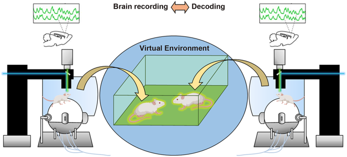
Synchronized dual microscopes connected via a shared virtual reality allow researchers to study neural dynamics and circuit mechanisms underlying social encounters and interactions of mice in a virtual world.
-
(3)
To take full advantage of our knowledge about the circuit mechanisms of social behavior, it is important to pursue not only pharmaceutical interventions but also non-pharmaceutical therapies. How can we establish circuit-based cures for disorders that affect social functioning? Despite its invasiveness, deep brain stimulation through implanted electrodes that directly intervene in pathological neural circuits has been applied to treat various neurological and neuropsychiatric disorders [155]. For the ASD population, non-invasive brain stimulation techniques such as transcranial magnetic stimulation and transcranial direct current stimulation have been used for treatment and rehabilitation, and meta-analyses of studies identify some beneficial effects on symptoms, including the social domain [156,157,158]. However, the efficacy, specificity, and safety of the current methods still face technical challenges. Next-generation non-invasive or minimally invasive neuromodulation technologies that use electric, optical, magnetic, and acoustic signals may offer a prospect for the clinical applications of circuit-specific brain stimulation therapy [159].

Humans can perform the most complex level of social behavior, and the action of brain networks that support such outstanding quality is extraordinarily intricate. Insights gained from future research using rodents as a model and their extension to explore principles shared with humans will continue to advance our understanding of the biological underpinnings of social behavior and associated deleterious changes. This endeavor will eventually lead to the development of circuit-based therapies for relevant disorders.
References
Chen P, Hong W. Neural circuit mechanisms of social behavior. Neuron. 2018;98:16–30.
Anderson DJ. Circuit modules linking internal states and social behaviour in flies and mice. Nat Rev Neurosci. 2016;17:692–704.
Wei D, Talwar V, Lin D. Neural circuits of social behaviors: innate yet flexible. Neuron. 2021;109:1600–20.
Wu YE, Hong W. Neural basis of prosocial behavior. Trends Neurosci. 2022;45:749–62.
Miura I, Overton ETN, Nakai N, Kawamata T, Sato M, Takumi T. Imaging the neural circuit basis of social behavior: insights from mouse and human studies. Neurol Med Chir (Tokyo). 2020;60:429–38.
Takumi T, Tamada K, Hatanaka F, Nakai N, Bolton PF. Behavioral neuroscience of autism. Neurosci Biobehav Rev. 2020;110:60–76.
Nakai N, Takumi T, Nakai J, Sato M. Common defects of spine dynamics and circuit function in neurodevelopmental disorders: a systematic review of findings from in vivo optical imaging of mouse models. Front Neurosci. 2018;12:412.
Huguet G, Ey E, Bourgeron T. The genetic landscapes of autism spectrum disorders. Annu Rev Genom Hum Genet. 2013;14:191–213.
Takumi T, Tamada K. CNV biology in neurodevelopmental disorders. Curr Opin Neurobiol. 2018;48:183–92.
Kim SW, Kim M, Shin HS. Affective empathy and prosocial behavior in rodents. Curr Opin Neurobiol. 2021;68:181–9.
Ko J. Neuroanatomical substrates of rodent social behavior: the medial prefrontal cortex and its projection patterns. Front Neural Circuits. 2017;11:41.
Amodio DM, Frith CD. Meeting of minds: the medial frontal cortex and social cognition. Nat Rev Neurosci. 2006;7:268–77.
Klein-Flügge MC, Bongioanni A, Rushworth MFS. Medial and orbital frontal cortex in decision-making and flexible behavior. Neuron. 2022;110:2743–70.
Yizhar O, Levy DR. The social dilemma: prefrontal control of mammalian sociability. Curr Opin Neurobiol. 2021;68:67–75.
Lee E, Rhim I, Lee JW, Ghim JW, Lee S, Kim E, et al. Enhanced neuronal activity in the medial prefrontal cortex during social approach behavior. J Neurosci. 2016;36:6926–36.
Yizhar O, Fenno LE, Prigge M, Schneider F, Davidson TJ, O’Shea DJ, et al. Neocortical excitation/inhibition balance in information processing and social dysfunction. Nature. 2011;477:171–8.
Liang B, Zhang L, Barbera G, Fang W, Zhang J, Chen X, et al. Distinct and dynamic ON and OFF neural ensembles in the prefrontal cortex code social exploration. Neuron. 2018;100:700–14.
Murugan M, Jang HJ, Park M, Miller EM, Cox J, Taliaferro JP, et al. Combined social and spatial coding in a descending projection from the prefrontal cortex. Cell. 2017;171:1663–77.
Huang WC, Zucca A, Levy J, Page DT. Social behavior is modulated by valence-encoding mPFC-amygdala sub-circuitry. Cell Rep. 2020;32:107899.
Felix-Ortiz AC, Burgos-Robles A, Bhagat ND, Leppla CA, Tye KM. Bidirectional modulation of anxiety-related and social behaviors by amygdala projections to the medial prefrontal cortex. Neuroscience. 2016;321:197–209.
Kuga N, Abe R, Takano K, Ikegaya Y, Sasaki T. Prefrontal-amygdalar oscillations related to social behavior in mice. eLife. 2022;11:e78428.
Liu L, Xu H, Wang J, Li J, Tian Y, Zheng J, et al. Cell type-differential modulation of prefrontal cortical GABAergic interneurons on low gamma rhythm and social interaction. Sci Adv. 2020;6:eaay4073.
Kim Y, Venkataraju KU, Pradhan K, Mende C, Taranda J, Turaga SC, et al. Mapping social behavior-induced brain activation at cellular resolution in the mouse. Cell Rep. 2015;10:292–305.
Nakajima M, Görlich A, Heintz N. Oxytocin modulates female sociosexual behavior through a specific class of prefrontal cortical interneurons. Cell. 2014;159:295–305.
Yamamuro K, Bicks LK, Leventhal MB, Kato D, Im S, Flanigan ME, et al. A prefrontal-paraventricular thalamus circuit requires juvenile social experience to regulate adult sociability in mice. Nat Neurosci. 2020;23:1240–52.
Park G, Ryu C, Kim S, Jeong SJ, Koo JW, Lee YS, et al. Social isolation impairs the prefrontal-nucleus accumbens circuit subserving social recognition in mice. Cell Rep. 2021;35:109104.
Benekareddy M, Stachniak TJ, Bruns A, Knoflach F, von Kienlin M, Künnecke B, et al. Identification of a corticohabenular circuit regulating socially directed behavior. Biol Psychiatry. 2018;83:607–17.
Yang Y, Wang H, Hu J, Hu H. Lateral habenula in the pathophysiology of depression. Curr Opin Neurobiol. 2018;48:90–6.
Kaminska B, Caballero JP, Moorman DE. Integration of value and action in medial prefrontal neural systems. Int Rev Neurobiol. 2021;158:57–82.
Gangopadhyay P, Chawla M, Dal Monte O, Chang SWC. Prefrontal-amygdala circuits in social decision-making. Nat Neurosci. 2021;24:5–18.
Jiang YH, Ehlers MD. Modeling autism by SHANK gene mutations in mice. Neuron. 2013;78:8–27.
Kim S, Kim YE, Song I, Ujihara Y, Kim N, Jiang YH, et al. Neural circuit pathology driven by Shank3 mutation disrupts social behaviors. Cell Rep. 2022;39:110906.
Li Y, Missig G, Finger BC, Landino SM, Alexander AJ, Mokler EL, et al. Maternal and early postnatal immune activation produce dissociable effects on neurotransmission in mPFC-amygdala circuits. J Neurosci. 2018;38:3358–72.
DeSpenza T Jr., Carlson M, Panchagnula S, Robert S, Duy PQ, Mermin-Bunnell N, et al. PTEN mutations in autism spectrum disorder and congenital hydrocephalus: developmental pleiotropy and therapeutic targets. Trends Neurosci. 2021;44:961–76.
Huang WC, Chen Y, Page DT. Hyperconnectivity of prefrontal cortex to amygdala projections in a mouse model of macrocephaly/autism syndrome. Nat Commun. 2016;7:13421.
Molosh AI, Johnson PL, Spence JP, Arendt D, Federici LM, Bernabe C, et al. Social learning and amygdala disruptions in Nf1 mice are rescued by blocking p21-activated kinase. Nat Neurosci. 2014;17:1583–90.
Nakai N, Otsuka S, Myung J, Takumi T. Autism spectrum disorder model mice: focus on copy number variation and epigenetics. Sci China Life Sci. 2015;58:976–84.
Qin L, Ma K, Wang ZJ, Hu Z, Matas E, Wei J, et al. Social deficits in Shank3-deficient mouse models of autism are rescued by histone deacetylase (HDAC) inhibition. Nat Neurosci. 2018;21:564–75.
van Heukelum S, Mars RB, Guthrie M, Buitelaar JK, Beckmann CF, Tiesinga PHE, et al. Where is cingulate cortex? A cross-species view. Trends Neurosci. 2020;43:285–99.
Apps MA, Rushworth MF, Chang SW. The anterior cingulate gyrus and social cognition: tracking the motivation of others. Neuron. 2016;90:692–707.
Chang SW, Gariépy JF, Platt ML. Neuronal reference frames for social decisions in primate frontal cortex. Nat Neurosci. 2013;16:243–50.
Amir N, Klumpp H, Elias J, Bedwell JS, Yanasak N, Miller LS. Increased activation of the anterior cingulate cortex during processing of disgust faces in individuals with social phobia. Biol Psychiatry. 2005;57:975–81.
Klumpp H, Fitzgerald JM, Kinney KL, Kennedy AE, Shankman SA, Langenecker SA, et al. Predicting cognitive behavioral therapy response in social anxiety disorder with anterior cingulate cortex and amygdala during emotion regulation. Neuroimage Clin. 2017;15:25–34.
de Waal FBM, Preston SD. Mammalian empathy: behavioural manifestations and neural basis. Nat Rev Neurosci. 2017;18:498–509.
Carcea I, Froemke RC. Biological mechanisms for observational learning. Curr Opin Neurobiol. 2019;54:178–85.
Keum S, Shin HS. Neural basis of observational fear learning: a potential model of affective empathy. Neuron. 2019;104:78–86.
Jeon D, Kim S, Chetana M, Jo D, Ruley HE, Lin SY, et al. Observational fear learning involves affective pain system and Cav1.2 Ca2+ channels in ACC. Nat Neurosci. 2010;13:482–8.
Allsop SA, Wichmann R, Mills F, Burgos-Robles A, Chang CJ, Felix-Ortiz AC, et al. Corticoamygdala transfer of socially derived information gates observational learning. Cell. 2018;173:1329–42.
Kim SW, Kim M, Baek J, Latchoumane CF, Gangadharan G, Yoon Y, et al. Hemispherically lateralized rhythmic oscillations in the cingulate-amygdala circuit drive affective empathy in mice. Neuron. 2023;111:418–29.
Smith ML, Asada N, Malenka RC. Anterior cingulate inputs to nucleus accumbens control the social transfer of pain and analgesia. Science. 2021;371:153–9.
Guo B, Chen J, Chen Q, Ren K, Feng D, Mao H, et al. Anterior cingulate cortex dysfunction underlies social deficits in Shank3 mutant mice. Nat Neurosci. 2019;22:1223–34.
Gogolla N. The insular cortex. Curr Biol. 2017;27:R580–6.
Livneh Y, Andermann ML. Cellular activity in insular cortex across seconds to hours: sensations and predictions of bodily states. Neuron. 2021;109:3576–93.
Allen GV, Saper CB, Hurley KM, Cechetto DF. Organization of visceral and limbic connections in the insular cortex of the rat. J Comp Neurol. 1991;311:1–16.
Shi CJ, Cassell MD. Cortical, thalamic, and amygdaloid connections of the anterior and posterior insular cortices. J Comp Neurol. 1998;399:440–68.
Gogolla N, Takesian AE, Feng G, Fagiolini M, Hensch TK. Sensory integration in mouse insular cortex reflects GABA circuit maturation. Neuron. 2014;83:894–905.
Rodgers KM, Benison AM, Klein A, Barth DS. Auditory, somatosensory, and multisensory insular cortex in the rat. Cereb Cortex. 2008;18:2941–51.
Klein AS, Dolensek N, Weiand C, Gogolla N. Fear balance is maintained by bodily feedback to the insular cortex in mice. Science. 2021;374:1010–5.
Koren T, Yifa R, Amer M, Krot M, Boshnak N, Ben-Shaanan TL, et al. Insular cortex neurons encode and retrieve specific immune responses. Cell. 2021;184:5902–15.
Jasmin L, Rabkin SD, Granato A, Boudah A, Ohara PT. Analgesia and hyperalgesia from GABA-mediated modulation of the cerebral cortex. Nature. 2003;424:316–20.
Tan LL, Pelzer P, Heinl C, Tang W, Gangadharan V, Flor H, et al. A pathway from midcingulate cortex to posterior insula gates nociceptive hypersensitivity. Nat Neurosci. 2017;20:1591–601.
Chen X, Gabitto M, Peng Y, Ryba NJ, Zuker CS. A gustotopic map of taste qualities in the mammalian brain. Science. 2011;333:1262–6.
Fletcher ML, Ogg MC, Lu L, Ogg RJ, Boughter JD Jr. Overlapping representation of primary tastes in a defined region of the gustatory cortex. J Neurosci. 2017;37:7595–605.
Chen K, Kogan JF, Fontanini A. Spatially distributed representation of taste quality in the gustatory insular cortex of behaving mice. Curr Biol. 2021;31:247–56.
Lavi K, Jacobson GA, Rosenblum K, Lüthi A. Encoding of conditioned taste aversion in cortico-amygdala circuits. Cell Rep. 2018;24:278–83.
Zhu J, Cheng Q, Chen Y, Fan H, Han Z, Hou R, et al. Transient delay-period activity of agranular insular cortex controls working memory maintenance in learning novel tasks. Neuron. 2020;105:934–46.
Yiannakas A, Kolatt Chandran S, Kayyal H, Gould N, Khamaisy M, Rosenblum K. Parvalbumin interneuron inhibition onto anterior insula neurons projecting to the basolateral amygdala drives aversive taste memory retrieval. Curr Biol. 2021;31:2770–84.
Dolensek N, Gehrlach DA, Klein AS, Gogolla N. Facial expressions of emotion states and their neuronal correlates in mice. Science. 2020;368:89–94.
Deng H, Xiao X, Yang T, Ritola K, Hantman A, Li Y, et al. A genetically defined insula-brainstem circuit selectively controls motivational vigor. Cell. 2021;184:6344–60.
Wang L, Gillis-Smith S, Peng Y, Zhang J, Chen X, Salzman CD, et al. The coding of valence and identity in the mammalian taste system. Nature. 2018;558:127–31.
Livneh Y, Ramesh RN, Burgess CR, Levandowski KM, Madara JC, Fenselau H, et al. Homeostatic circuits selectively gate food cue responses in insular cortex. Nature. 2017;546:611–6.
Livneh Y, Sugden AU, Madara JC, Essner RA, Flores VI, Sugden LA, et al. Estimation of current and future physiological states in insular cortex. Neuron. 2020;105:1094–111.
Gehrlach DA, Dolensek N, Klein AS, Roy Chowdhury R, Matthys A, Junghänel M, et al. Aversive state processing in the posterior insular cortex. Nat Neurosci. 2019;22:1424–37.
Wu Y, Chen C, Chen M, Qian K, Lv X, Wang H, et al. The anterior insular cortex unilaterally controls feeding in response to aversive visceral stimuli in mice. Nat Commun. 2020;11:640.
Miura I, Sato M, Overton ETN, Kunori N, Nakai J, Kawamata T, et al. Encoding of social exploration by neural ensembles in the insular cortex. PLoS Biol. 2020;18:e3000584.
Ramos-Prats A, Paradiso E, Castaldi F, Sadeghi M, Mir MY, Hörtnagl H, et al. VIP-expressing interneurons in the anterior insular cortex contribute to sensory processing to regulate adaptive behavior. Cell Rep. 2022;39:110893.
Rogers-Carter MM, Varela JA, Gribbons KB, Pierce AF, McGoey MT, Ritchey M, et al. Insular cortex mediates approach and avoidance responses to social affective stimuli. Nat Neurosci. 2018;21:404–14.
Rogers-Carter MM, Djerdjaj A, Gribbons KB, Varela JA, Christianson JP. Insular cortex projections to nucleus accumbens core mediate social approach to stressed juvenile rats. J Neurosci. 2019;39:8717–29.
Zhang MM, Geng AQ, Chen K, Wang J, Wang P, Qiu XT, et al. Glutamatergic synapses from the insular cortex to the basolateral amygdala encode observational pain. Neuron. 2022;110:1993–2008.
Craig AD. How do you feel-now? The anterior insula and human awareness. Nat Rev Neurosci. 2009;10:59–70.
Uddin LQ. Salience processing and insular cortical function and dysfunction. Nat Rev Neurosci. 2015;16:55–61.
Nomi JS, Molnar-Szakacs I, Uddin LQ. Insular function in autism: update and future directions in neuroimaging and interventions. Prog Neuropsychopharmacol Biol Psychiatry. 2019;89:412–26.
Mow JL, Gandhi A, Fulford D. Imaging the “social brain” in schizophrenia: a systematic review of neuroimaging studies of social reward and punishment. Neurosci Biobehav Rev. 2020;118:704–22.
Gehrlach DA, Weiand C, Gaitanos TN, Cho E, Klein AS, Hennrich AA, et al. A whole-brain connectivity map of mouse insular cortex. eLife. 2020;9:e55585.
O’Connell LA, Hofmann HA. The vertebrate mesolimbic reward system and social behavior network: a comparative synthesis. J Comp Neurol. 2011;519:3599–639.
O’Connell LA, Hofmann HA. Evolution of a vertebrate social decision-making network. Science. 2012;336:1154–7.
Newman SW. The medial extended amygdala in male reproductive behavior. A node in the mammalian social behavior network. Ann N. Y Acad Sci. 1999;877:242–57.
Rogers-Carter MM, Christianson JP. An insular view of the social decision-making network. Neurosci Biobehav Rev. 2019;103:119–32.
Bird CW, Barto D, Magcalas CM, Rodriguez CI, Donaldson T, Davies S, et al. Ifenprodil infusion in agranular insular cortex alters social behavior and vocalizations in rats exposed to moderate levels of ethanol during prenatal development. Behav Brain Res. 2017;320:1–11.
Lockwood PL. The anatomy of empathy: vicarious experience and disorders of social cognition. Behav Brain Res. 2016;311:255–66.
Langford DJ, Crager SE, Shehzad Z, Smith SB, Sotocinal SG, Levenstadt JS, et al. Social modulation of pain as evidence for empathy in mice. Science. 2006;312:1967–70.
Zaniboni CR, Pelarin V, Baptista-de-Souza D, Canto-de-Souza A. Empathy for pain: insula inactivation and systemic treatment with midazolam reverses the hyperalgesia induced by cohabitation with a pair in chronic pain condition. Front Behav Neurosci. 2018;12:278.
Burkett JP, Andari E, Johnson ZV, Curry DC, de Waal FB, Young LJ. Oxytocin-dependent consolation behavior in rodents. Science. 2016;351:375–8.
Matsumoto M, Yoshida M, Jayathilake BW, Inutsuka A, Nishimori K, Takayanagi Y, et al. Indispensable role of the oxytocin receptor for allogrooming toward socially distressed cage mates in female mice. J Neuroendocrinol. 2021;33:e12980.
Wu YE, Dang J, Kingsbury L, Zhang M, Sun F, Hu RK, et al. Neural control of affiliative touch in prosocial interaction. Nature. 2021;599:262–7.
Ben-Ami Bartal I, Decety J, Mason P. Empathy and pro-social behavior in rats. Science. 2011;334:1427–30.
Cox SS, Kearns AM, Woods SK, Brown BJ, Brown SJ, Reichel CM. The role of the anterior insular during targeted helping behavior in male rats. Sci Rep. 2022;12:3315.
Singer T, Seymour B, O’Doherty J, Kaube H, Dolan RJ, Frith CD. Empathy for pain involves the affective but not sensory components of pain. Science. 2004;303:1157–62.
Lamm C, Decety J, Singer T. Meta-analytic evidence for common and distinct neural networks associated with directly experienced pain and empathy for pain. Neuroimage. 2011;54:2492–502.
Wicker B, Keysers C, Plailly J, Royet JP, Gallese V, Rizzolatti G. Both of us disgusted in my insula: the common neural basis of seeing and feeling disgust. Neuron. 2003;40:655–64.
Gu X, Gao Z, Wang X, Liu X, Knight RT, Hof PR, et al. Anterior insular cortex is necessary for empathetic pain perception. Brain. 2012;135:2726–35.
Aoki Y, Yahata N, Watanabe T, Takano Y, Kawakubo Y, Kuwabara H, et al. Oxytocin improves behavioural and neural deficits in inferring others’ social emotions in autism. Brain. 2014;137:3073–86.
Djerdjaj A, Ng AJ, Rieger NS, Christianson JP. The basolateral amygdala to posterior insular cortex tract is necessary for social interaction with stressed juvenile rats. Behav Brain Res. 2022;435:114050.
McFarlane HG, Kusek GK, Yang M, Phoenix JL, Bolivar VJ, Crawley JN. Autism-like behavioral phenotypes in BTBR T+tf/J mice. Genes Brain Behav. 2008;7:152–63.
Froemke RC, Young LJ. Oxytocin, neural plasticity, and social behavior. Annu Rev Neurosci. 2021;44:359–81.
Knobloch HS, Charlet A, Hoffmann LC, Eliava M, Khrulev S, Cetin AH, et al. Evoked axonal oxytocin release in the central amygdala attenuates fear response. Neuron. 2012;73:553–66.
Son S, Manjila SB, Newmaster KT, Wu YT, Vanselow DJ, Ciarletta M, et al. Whole-brain wiring diagram of oxytocin system in adult mice. J Neurosci. 2022;42:5021–33.
Ferguson JN, Young LJ, Hearn EF, Matzuk MM, Insel TR, Winslow JT. Social amnesia in mice lacking the oxytocin gene. Nat Genet. 2000;25:284–8.
Takayanagi Y, Yoshida M, Bielsky IF, Ross HE, Kawamata M, Onaka T, et al. Pervasive social deficits, but normal parturition, in oxytocin receptor-deficient mice. Proc Natl Acad Sci USA. 2005;102:16096–101.
Ferguson JN, Aldag JM, Insel TR, Young LJ. Oxytocin in the medial amygdala is essential for social recognition in the mouse. J Neurosci. 2001;21:8278–85.
Raam T, McAvoy KM, Besnard A, Veenema AH, Sahay A. Hippocampal oxytocin receptors are necessary for discrimination of social stimuli. Nat Commun. 2017;8:2001.
Marlin BJ, Mitre M, D’Amour JA, Chao MV, Froemke RC. Oxytocin enables maternal behaviour by balancing cortical inhibition. Nature. 2015;520:499–504.
Young LJ, Lim MM, Gingrich B, Insel TR. Cellular mechanisms of social attachment. Horm Behav. 2001;40:133–8.
Hung LW, Neuner S, Polepalli JS, Beier KT, Wright M, Walsh JJ, et al. Gating of social reward by oxytocin in the ventral tegmental area. Science. 2017;357:1406–11.
Dölen G, Darvishzadeh A, Huang KW, Malenka RC. Social reward requires coordinated activity of nucleus accumbens oxytocin and serotonin. Nature. 2013;501:179–84.
Salgado S, Kaplitt MG. The nucleus accumbens: a comprehensive review. Stereotact Funct Neurosurg. 2015;93:75–93.
Gunaydin LA, Grosenick L, Finkelstein JC, Kauvar IV, Fenno LE, Adhikari A, et al. Natural neural projection dynamics underlying social behavior. Cell. 2014;157:1535–51.
Krishnan V, Han MH, Graham DL, Berton O, Renthal W, Russo SJ, et al. Molecular adaptations underlying susceptibility and resistance to social defeat in brain reward regions. Cell. 2007;131:391–404.
Chaudhury D, Walsh JJ, Friedman AK, Juarez B, Ku SM, Koo JW, et al. Rapid regulation of depression-related behaviours by control of midbrain dopamine neurons. Nature. 2013;493:532–6.
Muir J, Lorsch ZS, Ramakrishnan C, Deisseroth K, Nestler EJ, Calipari ES, et al. In vivo fiber photometry reveals signature of future stress susceptibility in nucleus accumbens. Neuropsychopharmacology. 2018;43:255–63.
Brumback AC, Ellwood IT, Kjaerby C, Iafrati J, Robinson S, Lee AT, et al. Identifying specific prefrontal neurons that contribute to autism-associated abnormalities in physiology and social behavior. Mol Psychiatry. 2018;23:2078–89.
Challis C, Beck SG, Berton O. Optogenetic modulation of descending prefrontocortical inputs to the dorsal raphe bidirectionally bias socioaffective choices after social defeat. Front Behav Neurosci. 2014;8:43.
Cavalcante LES, Zinn CG, Schmidt SD, Saenger BF, Ferreira FF, Furini CRG, et al. Modulation of the storage of social recognition memory by neurotransmitter systems in the insular cortex. Behav Brain Res. 2017;334:129–34.
Nakai N, Nagano M, Saitow F, Watanabe Y, Kawamura Y, Kawamoto A, et al. Serotonin rebalances cortical tuning and behavior linked to autism symptoms in 15q11-13 CNV mice. Sci Adv. 2017;3:e1603001.
Walsh JJ, Christoffel DJ, Heifets BD, Ben-Dor GA, Selimbeyoglu A, Hung LW, et al. 5-HT release in nucleus accumbens rescues social deficits in mouse autism model. Nature. 2018;560:589–94.
Isles AR, Ingason A, Lowther C, Walters J, Gawlick M, Stöber G, et al. Parental origin of interstitial duplications at 15q11.2-q13.3 in Schizophrenia and neurodevelopmental disorders. PLoS Genet. 2016;12:e1005993.
Nakatani J, Tamada K, Hatanaka F, Ise S, Ohta H, Inoue K, et al. Abnormal behavior in a chromosome-engineered mouse model for human 15q11-13 duplication seen in autism. Cell. 2009;137:1235–46.
Tamada K, Tomonaga S, Hatanaka F, Nakai N, Takao K, Miyakawa T, et al. Decreased exploratory activity in a mouse model of 15q duplication syndrome; implications for disturbance of serotonin signaling. PLoS One. 2010;5:e15126.
Chung C, Shin W, Kim E. Early and late corrections in mouse models of autism spectrum disorder. Biol Psychiatry. 2022;91:934–44.
Yin X, Jones N, Yang J, Asraoui N, Mathieu ME, Cai L, et al. Delayed motor learning in a 16p11.2 deletion mouse model of autism is rescued by locus coeruleus activation. Nat Neurosci. 2021;24:646–57.
Zhang HF, Dai YC, Wu J, Jia MX, Zhang JS, Shou XJ, et al. Plasma oxytocin and Arginine-Vasopressin levels in children with autism spectrum disorder in China: associations with symptoms. Neurosci Bull. 2016;32:423–32.
Rodrigues SM, Saslow LR, Garcia N, John OP, Keltner D. Oxytocin receptor genetic variation relates to empathy and stress reactivity in humans. Proc Natl Acad Sci USA. 2009;106:21437–41.
LoParo D, Waldman ID. The oxytocin receptor gene (OXTR) is associated with autism spectrum disorder: a meta-analysis. Mol Psychiatry. 2015;20:640–6.
Jin D, Liu HX, Hirai H, Torashima T, Nagai T, Lopatina O, et al. CD38 is critical for social behaviour by regulating oxytocin secretion. Nature. 2007;446:41–45.
Fujima S, Yamaga R, Minami H, Mizuno S, Shinoda Y, Sadakata T, et al. CAPS2 deficiency impairs the release of the social peptide oxytocin, as well as oxytocin-associated social behavior. J Neurosci. 2021;41:4524–35.
Peñagarikano O, Lázaro MT, Lu XH, Gordon A, Dong H, Lam HA, et al. Exogenous and evoked oxytocin restores social behavior in the Cntnap2 mouse model of autism. Sci Transl Med. 2015;7:271ra278.
Sgritta M, Dooling SW, Buffington SA, Momin EN, Francis MB, Britton RA, et al. Mechanisms underlying microbial-mediated changes in social behavior in mouse models of autism spectrum disorder. Neuron. 2019;101:246–59.
Harony-Nicolas H, Kay M, du Hoffmann J, Klein ME, Bozdagi-Gunal O, Riad M, et al. Oxytocin improves behavioral and electrophysiological deficits in a novel Shank3-deficient rat. eLife. 2017;6:e18904.
Lewis EM, Stein-O’Brien GL, Patino AV, Nardou R, Grossman CD, Brown M, et al. Parallel social information processing circuits are differentially impacted in autism. Neuron. 2020;108:659–75.
Hörnberg H, Pérez-Garci E, Schreiner D, Hatstatt-Burklé L, Magara F, Baudouin S, et al. Rescue of oxytocin response and social behaviour in a mouse model of autism. Nature. 2020;584:252–6.
Bariselli S, Tzanoulinou S, Glangetas C, Prévost-Solié C, Pucci L, Viguié J, et al. SHANK3 controls maturation of social reward circuits in the VTA. Nat Neurosci. 2016;19:926–34.
Choe KY, Bethlehem RAI, Safrin M, Dong H, Salman E, Li Y, et al. Oxytocin normalizes altered circuit connectivity for social rescue of the Cntnap2 knockout mouse. Neuron. 2022;110:795–808.
Renier N, Adams EL, Kirst C, Wu Z, Azevedo R, Kohl J, et al. Mapping of brain activity by automated volume analysis of immediate early genes. Cell. 2016;165:1789–802.
Johnson ZV, Walum H, Xiao Y, Riefkohl PC, Young LJ. Oxytocin receptors modulate a social salience neural network in male prairie voles. Horm Behav. 2017;87:16–24.
Bosch OJ, Dabrowska J, Modi ME, Johnson ZV, Keebaugh AC, Barrett CE, et al. Oxytocin in the nucleus accumbens shell reverses CRFR2-evoked passive stress-coping after partner loss in monogamous male prairie voles. Psychoneuroendocrinology. 2016;64:66–78.
Mague SD, Talbot A, Blount C, Walder-Christensen KK, Duffney LJ, Adamson E, et al. Brain-wide electrical dynamics encode individual appetitive social behavior. Neuron. 2022;110:1728–41.
Nakai N, Sato M, Yamashita O, Sekine Y, Fu X, Nakai J, et al. Virtual reality-based real-time imaging reveals abnormal cortical dynamics during behavioral transitions in a mouse model of autism. Cell Rep. 2023;42:112258.
Ota K, Oisi Y, Suzuki T, Ikeda M, Ito Y, Ito T, et al. Fast, cell-resolution, contiguous-wide two-photon imaging to reveal functional network architectures across multi-modal cortical areas. Neuron. 2021;109:1810–24.
Lecoq J, Savall J, Vučinić D, Grewe BF, Kim H, Li JZ, et al. Visualizing mammalian brain area interactions by dual-axis two-photon calcium imaging. Nat Neurosci. 2014;17:1825–9.
Kingsbury L, Hong W. A multi-brain framework for social interaction. Trends Neurosci. 2020;43:651–66.
Kingsbury L, Huang S, Wang J, Gu K, Golshani P, Wu YE, et al. Correlated neural activity and encoding of behavior across brains of socially interacting animals. Cell. 2019;178:429–46.
Zhang W, Yartsev MM. Correlated neural activity across the brains of socially interacting bats. Cell. 2019;178:413–28.
Usmani SS, Sharath M, Mehendale M. Future of mental health in the metaverse. Gen Psychiatr. 2022;35:e100825.
Calabrò RS, Cerasa A, Ciancarelli I, Pignolo L, Tonin P, Iosa M, et al. The arrival of the metaverse in neurorehabilitation: fact, fake or vision? Biomedicines. 2022;10:2602.
Lozano AM, Lipsman N, Bergman H, Brown P, Chabardes S, Chang JW, et al. Deep brain stimulation: current challenges and future directions. Nat Rev Neurol. 2019;15:148–60.
Finisguerra A, Borgatti R, Urgesi C. Non-invasive brain stimulation for the rehabilitation of children and adolescents with neurodevelopmental disorders: a systematic review. Front Psychol. 2019;10:135.
García-González S, Lugo-Marín J, Setien-Ramos I, Gisbert-Gustemps L, Arteaga-Henríquez G, Díez-Villoria E, et al. Transcranial direct current stimulation in Autism Spectrum Disorder: a systematic review and meta-analysis. Eur Neuropsychopharmacol. 2021;48:89–109.
Rosson S, de Filippis R, Croatto G, Collantoni E, Pallottino S, Guinart D, et al. Brain stimulation and other biological non-pharmacological interventions in mental disorders: an umbrella review. Neurosci Biobehav Rev. 2022;139:104743.
Liu X, Qiu F, Hou L, Wang X. Review of noninvasive or minimally invasive deep brain stimulation. Front Behav Neurosci. 2021;15:820017.
Acknowledgements
We thank Yu Ohmura for his valuable comments on this manuscript.
Funding
The authors receive financial support from: The KAKENHI from JSPS (20H03550 and 23H02668) and Taiju Life Social Welfare Foundation to MS; The KAKENHI from JSPS (23K14673 and 23H04138) to NN; The KAKENHI from JSPS (16H06316, 16H06463, 21H00202, 21H04813, and 23H04233), Japan Agency for Medical Research and Development (JP21wm0425011), Japan Science and Technology Agency (JPMJMS2299 and JPMJMS229B), Intramural Research Grant (30-9) for Neurological and Psychiatric Disorders of NCNP, The Takeda Science Foundation, Research Foundation for Opto-Science and Technology, Taiju Life Social Welfare Foundation, The Naito Foundation, The Tokumori Yasumoto Memorial Trust for Researches on Tuberous Sclerosis Complex and Related Rare Neurological Diseases to TT, and the Canada Research Chairs Program to KYC. Open access funding provided by Kobe University.
Author information
Authors and Affiliations
Contributions
MS, NN, SF, KYC, and TT wrote and edited the manuscript.
Corresponding author
Ethics declarations
Competing interests
The authors declare no competing interests.
Additional information
Publisher’s note Springer Nature remains neutral with regard to jurisdictional claims in published maps and institutional affiliations.
Rights and permissions
Open Access This article is licensed under a Creative Commons Attribution 4.0 International License, which permits use, sharing, adaptation, distribution and reproduction in any medium or format, as long as you give appropriate credit to the original author(s) and the source, provide a link to the Creative Commons licence, and indicate if changes were made. The images or other third party material in this article are included in the article’s Creative Commons licence, unless indicated otherwise in a credit line to the material. If material is not included in the article’s Creative Commons licence and your intended use is not permitted by statutory regulation or exceeds the permitted use, you will need to obtain permission directly from the copyright holder. To view a copy of this licence, visit http://creativecommons.org/licenses/by/4.0/.
About this article

Cite this article
Sato, M., Nakai, N., Fujima, S. et al. Social circuits and their dysfunction in autism spectrum disorder. Mol Psychiatry 28, 3194–3206 (2023). https://doi.org/10.1038/s41380-023-02201-0
Received:
Revised:
Accepted:
Published:
Issue Date:
DOI: https://doi.org/10.1038/s41380-023-02201-0
