


« Prev Next »

CDK
Multiple checkpoints in the eukaryotic cell cycle ensure that division occurs only after sufficient growth and faithful DNA replication, and only when favorable conditions exist. At each checkpoint, numerous proteins engage in a series of carefully coordinated biochemical reactions. This complexity allows for precise regulation of all steps in the cell cycle — and it is essential to preventing the devastating consequences of cell division gone awry (Figure 1).
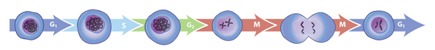
Figure 1: The sequence of eukaryotic cell cycle phases
Between each arrow, the cell passes through a particular cell cycle checkpoint.
© 2013 Nature Education All rights reserved. 
What Are Cyclin-Dependent Kinases?
Of the many proteins involved in cell cycle control, cyclin-dependent kinases (CDKs) are among the most important. CDKs are a family of multifunctional enzymes that can modify various protein substrates involved in cell cycle progression. Specifically, CDKs phosphorylate their substrates by transferring phosphate groups from ATP to specific stretches of amino acids in the substrates. Different types of eukaryotic cells contain different types and numbers of CDKs. For example, yeast have only a single CDK, whereas vertebrates have four different ones.
As their name suggests, CDKs require the presence of cyclins to become active. Cyclins are a family of proteins that have no enzymatic activity of their own but activate CDKs by binding to them. CDKs must also be in a particular phosphorylation state — with some sites phosphorylated and others dephosphorylated — in order for activation to occur. Correct phosphorylation depends on the action of other kinases and a second class of enzymes called phosphatases that are responsible for removing phosphate groups from proteins.
How Do CDKs Control the Cell Cycle?
All eukaryotes have multiple cyclins, each of which acts during a specific stage of the cell cycle. (In organisms with multiple CDKs, each CDK is paired with a specific cyclin.) All cyclins are named according to the stage at which they assemble with CDKs. Common classes of cyclins include G1-phase cyclins, G1/S-phase cyclins, S-phase cyclins, and M-phase cyclins. M-phase cyclins form M-CDK complexes and drive the cell's entry into mitosis; G1 cyclins form G1-CDK complexes and guide the cell's progress through the G1 phase; and so on.
All CDKs exist in similar amounts throughout the entire cell cycle. In contrast, cyclin manufacture and breakdown varies by stage — with cell cycle progression dependent on the synthesis of new cyclin molecules. Accordingly, cells synthesize G1- and G1/S-cyclins at different times during the G1 phase, and they produce M-cyclin molecules during the G2 phase (Figure 2). Cyclin degradation is equally important for progression through the cell cycle. Specific enzymes break down cyclins at defined times in the cell cycle. When cyclin levels decrease, the corresponding CDKs become inactive. Cell cycle arrest can occur if cyclins fail to degrade.
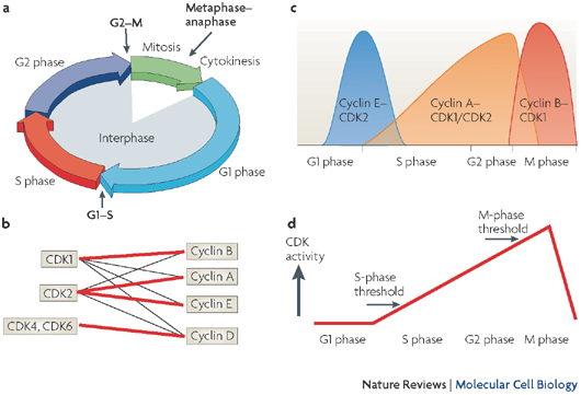
Figure 2: The classical and minimal models of cell cycle control
Where and when do cyclins act on the cell cycle? (A) Cycling cells undergo three major transitions during their cell cycle. The beginning of S phase is marked by the onset of DNA replication, the start of mitosis (M) is accompanied by breakdown of the nuclear envelope and chromosome condensation, whereas segregation of the sister chromatids marks the metaphase-to-anaphase transition. Cyclin-dependent kinases (CDKs) trigger the transition from G1 to S phase and from G2 to M phase by phosphorylating distinct sets of substrates. (B) CDK1 and CDK2 bind to multiple cyclins (cyclin types A, B, D and E), whereas CDK4 and CDK6 only partner D-type cyclins. Thick lines represent the preferred pairing for each kinase. (C) According to the classical model of cell cycle control, D-type cyclins and CDK4 or CDK6 regulate events in early G1 phase (not shown), cyclin E-CDK2 triggers S phase, cyclin A-CDK2 and cyclin A-CDK1 regulate the completion of S phase, and CDK1-cyclin B is responsible for mitosis. (D) Based on the results of cyclin and CDK-knockout studies, scientists have constructed a new threshold model of cell cycle control. Accordingly, either CDK1 or CDK2 bound to cyclin A is sufficient to control interphase, whereas cyclin B-CDK1 is essential to take cells into mitosis. The differences between interphase and mitotic CDKs are not necessarily due to substrate specificity, but are more likely a result of different localization and a higher activity threshold for mitosis than interphase.
© 2008 Nature Publishing Group Hochegger, H., Takeda, S., & Hunt, T. Cyclin-dependent kinases and cell-cycle transitions: does one fit all? Nature Reviews Molecular Cell Biology 9, 910-916 (2008). All rights reserved.
Which Proteins Do CDKs Modify?
Each
of the cyclin-CDK complexes in a cell modifies a specific group of protein
substrates. Proper phosphorylation of these substrates must occur at
particular
times in order for the cell cycle to continue. Because cyclin-CDK
complexes
recognize multiple substrates, they are able to coordinate the multiple
events
that occur during each phase of the cell cycle. For example, at the
beginning
of S phase, S-CDK catalyzes phosphorylation of the proteins that
initiate DNA
replication by allowing DNA replication complexes to form. Later, during
mitosis, M-CDKs phosphorylate a wide range of proteins. These include
condensin
proteins, which are essential for the extensive condensation of mitotic
chromosomes, and lamin proteins, which form a stabilizing network under
the
nuclear membrane that dissembles during mitosis. M-CDKs also influence
the
assembly of the mitotic spindle by phosphorylating proteins that
regulate
microtubule behavior. The net effect of these coordinated
phosphorylation
reactions is the accurate separation of chromosomes during mitosis.
Conclusion
The life cycle of a cell is a carefully regulated series of events orchestrated
by a
suite of enzymes and other proteins. The main regulatory components of
cell
cycle control are cyclins and CDKs. Depending on the presence and action
of
these proteins, the cell cycle can be speedy or slow, and it may even
halt
altogether.
eBooks
This page appears in the following eBook


