Thank you for visiting nature.com. You are using a browser version with limited support for CSS. To obtain
the best experience, we recommend you use a more up to date browser (or turn off compatibility mode in
Internet Explorer). In the meantime, to ensure continued support, we are displaying the site without styles
and JavaScript.
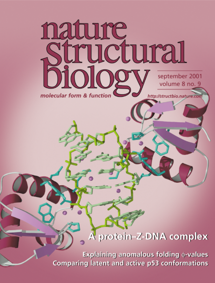
Crystal structure of ZαDLM (ribbon diagram) in complex with left-handed Z-DNA (yellow stick model). ZαDLM is a Z-DNA binding domain from a tumor-associated protein, DLM-1. This is the second structure reported for a Z-DNA–protein complex. DNA recognition is mediated by direct and water-mediated contacts (protein residues, light blue stick model; water molecules, purple spheres). The recognition mode is similar to that observed in the first Z-DNA–protein structure, suggesting that this family of proteins shares a common Z-DNA binding motif. See pages 761–765.
NMR analyses of variants of the p53 tumor suppressor protein that either contain or lack the C-terminus reveal that the C-terminus neither interacts with other regions of p53 nor has an impact on the conformation of the rest of the molecule. Nevertheless, the C-terminus is likely to be critical for regulation of p53 function.
Copper is an essential transition metal that plays a fundamental role in the biochemistry of all aerobic organisms. Recent work elucidating the structural mechanisms of copper delivery to superoxide dismutase provides insight into the cell biology of copper metabolism and serves as an example of how to understand the principles governing the incorporation of metals into proteins.
The first crystal structure of an actin molecule not associated with another protein has been solved. Remarkably, the most variable part of actin's structure is also the most conserved part of its sequence.
Unambiguous evidence for a glycosyl-enzyme intermediate on the lysozyme reaction pathway has recently been reported, finally settling what kind of mechanism this textbook enzyme uses.