Volume 9
-
No. 12 December 2013
Cover image supplied by Ms Susann Junker, Prof Ulf Muller-Ladner and Dr Elena Neumann from the Internal Medicine and Rheumatology, Justus-Liebig-University of Gieen, Germany. The image shows a formalin-fixed, decalcified and paraffin embedded grade 5 osteophyte from a patient with osteoarthritis. Masson's trichrome staining was used for visualizing areas of cartilage, connective tissue and osteoid within the osteophyte. The osteophyte contains a thick layer of cartilage, ossified remodeling zones and mineralized areas with dense osteoid. This staining was performed as part of a project to investigate osteophyte development in osteoarthritis, especially regarding the role of adipokines in this process.
-
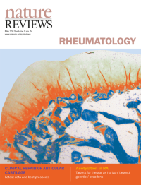
No. 11 November 2013
Cover image supplied by Ms Susann Junker, Prof Ulf Muller-Ladner and Dr Elena Neumann from the Internal Medicine and Rheumatology, Justus-Liebig-University of Gieen, Germany. The image shows a formalin-fixed, decalcified and paraffin embedded grade 5 osteophyte from a patient with osteoarthritis. Masson's trichrome staining was used for visualizing areas of cartilage, connective tissue and osteoid within the osteophyte. The osteophyte contains a thick layer of cartilage, ossified remodeling zones and mineralized areas with dense osteoid. This staining was performed as part of a project to investigate osteophyte development in osteoarthritis, especially regarding the role of adipokines in this process.
-
No. 10 October 2013
Cover image supplied by Ms Susann Junker, Prof Ulf Muller-Ladner and Dr Elena Neumann from the Internal Medicine and Rheumatology, Justus-Liebig-University of Gieen, Germany. The image shows a formalin-fixed, decalcified and paraffin embedded grade 5 osteophyte from a patient with osteoarthritis. Masson's trichrome staining was used for visualizing areas of cartilage, connective tissue and osteoid within the osteophyte. The osteophyte contains a thick layer of cartilage, ossified remodeling zones and mineralized areas with dense osteoid. This staining was performed as part of a project to investigate osteophyte development in osteoarthritis, especially regarding the role of adipokines in this process.
-
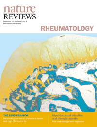
No. 9 September 2013
Cover image supplied by Ms Susann Junker, Prof Ulf Muller-Ladner and Dr Elena Neumann from the Internal Medicine and Rheumatology, Justus-Liebig-University of Gieen, Germany. The image shows a formalin-fixed, decalcified and paraffin embedded grade 5 osteophyte from a patient with osteoarthritis. Masson's trichrome staining was used for visualizing areas of cartilage, connective tissue and osteoid within the osteophyte. The osteophyte contains a thick layer of cartilage, ossified remodeling zones and mineralized areas with dense osteoid. This staining was performed as part of a project to investigate osteophyte development in osteoarthritis, especially regarding the role of adipokines in this process.