Volume 9
-
No. 12 December 2014
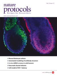
Peri-implantation embryogenesis is largely unexplored and probably the most enigmatic period of mouse development. The protocol by Bedzhov et al. describes how to support murine embryonic development beyond the blastocyst stage in vitro allowing direct observation of blastocyst to egg cylinder transition. The egg cylinder shown on the cover was recovered at E6.5. Oct4 (red) marks the epiblast. Eomes (green) is expressed in the posterior EPI, extraembryonic ectoderm and visceral endoderm. DAPI (blue) marks the nuclei.
-
No. 11 November 2014
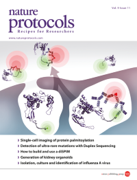
Metabolic bioorthogonal labeling of proteins within cells with an alkynyl analog of palmitic acid followed by click chemistry and an in situ proximity ligation assay enables the cellular imaging of palmitoylation in a protein of interest. The image depicts palmitoylated Wnt proteins (rendered from PDB file 4F0A) en route to secretion outside the cell. Based on the protocol by Xinxin Gao and Rami N. Hannoush. doi: 10.1038/nprot.2014.179. Image generated by Allison Bruce. Cover design by Jamel Wooten.
-
No. 10 October 2014
A population of bacterial genomes can be rapidly edited at many loci using multiplex automated genome engineering (MAGE; Gallagher et al. doi:10.1038/nprot.2014.082). As more MAGE cycles are performed, the most prevalent genotype shifts from being unedited to fully edited (colored bars), passing through a period of greater variability (black line). This process can be enhanced by co-selection MAGE (right). Cover design by Jamel Wooten.
-
No. 9 September 2014
Scanning electron microscopy image of an electrospun artificial trachea showing a fibrous architecture similar to that seen in the native rat trachea. Image taken from the protocol by Jungebluth et al. doi:10.1038/nprot.2014.149. Cover design by Jamel Wooten.
-
No. 8 August 2014
Fourier transform infrared (FTIR) spectroscopy can allow for the derivation of rich biochemical images in a label-free manner, which can be used in disease detection. This FTIR image is from a kidney biopsy, highlighting a glomerulus by CH2 asymmetric absorbance. Image by Vishal K Varma, Holly J Butler, Suman Setty, Francis L Martin, Michael J Walsh (University of Illinois at Chicago, USA; Lancaster University, UK), associated with the protocol by Baker et al. doi: 10.1038/nprot.2014.110.
-
No. 7 July 2014
Zebrafish larval positional preference within a well, analyzed with LSRtrack (Zhou et al. doi:10.1038/nprot.2014.094). Larvae tended to occupy the center of the well when exposed to bright light, whereas in darkness the same animals swam close to the walls of the well ('thigmotaxis').
-
No. 6 June 2014
Inner ear organoids can be produced from mouse embryonic stem cells grown under chemically defined conditions by following the protocol by Koehler and Hashino (doi:10.1038/nprot.2014.100). Two organoid variants develop; this modified image includes an embedded organoid fixed and stained with antibodies specific to cyclin D1 (red) and calretinin (green) 20 d after initiation of differentiation.
-
No. 5 May 2014
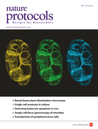
Maximum projection views of the same falsely colored live C. elegans embryo, labeled with a GFP cell membrane marker and imaged using Bessel beam structured illumination microscopy. Image adapted from the protocol by Gao et al. doi:10.1038/nprot.2014.087. Cover design by Jamel Wooten.
-
No. 4 April 2014
Haploid Arabidopsis plants are easy to generate and have small, narrow leaves. The protocol by Wijnker et al. (doi:10.1038/nprot.2014.049) describes how (haploid) chromosome substitution lines (CSLs) are generated from Arabidopsis heterozygotes during reverse breeding. These CSLs subsequently serve as parental lines to recreate the heterozygote.
-
No. 3 March 2014
The axolotl (Ambystoma mexicanum) is a very useful model organism for studying limb and spinal cord regeneration. The protocol by Khattak et al. (doi: 10.1038/nprot.2014.040) describes optimized axolotl husbandry, breeding, metamorphosis, transgenesis and tamoxifen-induced, Cre-mediated recombination.
-
No. 2 February 2014
3D cell motion on a sagittal section of a mid-gastrulation Xenopus laevis embryo. Red arrows designate maximum cell speed (∼4 μm min1): mesendodermal leading-edge runners (top center); endodermal wall of inflating archenteron (left); blastopore (bottom center). Note pronounced vegetal rotation on lower right and epibolic movement throughout the periphery. Image from the protocol by Moosmann et al. doi: 10.1038/nprot.2014.033. Cover design by Jamel Wooten.
-
No. 1 January 2014
On the cover is an image of a fly using its rear leg to clean itself during a grooming reflex. Fruit flies have many innate behaviors that allow neuroscientists to investigate how the hard-wired neural circuits underlying these behaviors self-assemble. Image from the protocol by Kays et al. doi: 10.1038/nprot.2013.157. Cover design by Jamel Wooten.