
Volume 7 Issue 5, May 2012
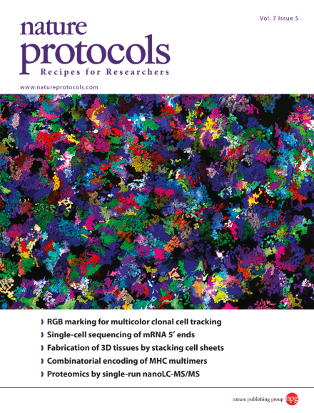
Microscopic photograph of RGB-marked 293T cells, 4 d after plating. Individual clones are distinguishable by specific colors resulting from the coexpression of three fluorescent proteins (red, green, blue) in highly characteristic amounts. Shown is an overlay of three fluorescence microscopy images obtained for the basic colors red, green and blue. Image taken from the protocol by Fehse et al. 10.1038/nprot.2012.026.
Protocol
-
Advertisement
