Thank you for visiting nature.com. You are using a browser version with limited support for CSS. To obtain
the best experience, we recommend you use a more up to date browser (or turn off compatibility mode in
Internet Explorer). In the meantime, to ensure continued support, we are displaying the site without styles
and JavaScript.
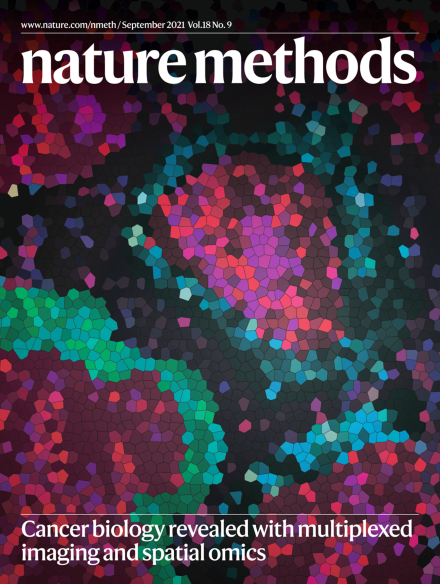
Cancer biology revealed with multiplexed imaging and spatial omics
Spatial omics and multiplexed imaging technologies are revealing cancer cell heterogeneity (red and purple cells) within a complex tumor microenvironment (blue, green and black cells).
Constructing a fair and accurate author list can be one of the most fraught aspects of manuscript publication. We provide some advice and resources for authors at all career levels.
Researchers use an in vitro assay in combination with a polymer model to study protein–DNA condensation and how condensates bring distant DNA elements into close proximity.
This Review describes spatial omics and multiplexed imaging technologies and their current and future impact in studying tumor heterogeneity and cancer biology.
This Review describes the state of the art in imaging extracellular vesicles in animals to study their release, biodistribution and uptake, and covers labeling strategies, microscopy methods and discoveries made in model organisms.
The LIVECell dataset comprises annotated phase-contrast images of over 1.6 million cells from different cell lines during growth from sparse seeding to confluence for improved training of deep learning-based models of image segmentation.
This analysis systematically evaluates cross-linking chemistry and chromatin fragmentation strategies commonly used in 3C assays and introduces an improved Hi-C protocol for detecting loops and compartments.
This work describes m6A-seq2, which utilizes multiplexed N6-methyladenosine (m6A) immunoprecipitation of pre-barcoded and pooled samples to reduce the technical variability and allows direct comparison of m6A at the site, gene and sample level.
This work describes nascent Ribo-Seq, which measures the speed of ribosome loading and polysome assembly on endogenous mRNA through a combination of 4-thiouridine labeling and ribosome footprint sequencing.
Long-range CRISPR activation can be enhanced by concurrent recruitment of artificial TFs to the enhancer and promoter of a target gene. This CRISPR activation system can be employed to achieve allele-selective gene upregulation by differentially targeting single-nucleotide polymorphisms embedded in enhancers or other distally located sequences.
DECODE uses deep learning for localizing single emitters in high-density two-dimensional and three-dimensional single-molecule localization microscopy data. DECODE outperforms available methods and enables fast live-cell SMLM of dynamic processes.
Mitometer enables efficient, rapid, and accurate automated segmentation and tracking of mitochondria from time-lapse images. Mitometer performs well on diverse input images and can be used to monitor dynamic fission and fusion events.