Volume 18
-
No. 12 December 2017
The activated immune system monopolizes the pool of amino acids in blood, diverting them from the brain and thus restricting the synthesis of serotonin and dopamine. Fagarasan and colleagues (p 1342; News and Views by Boussiotis p 1281) show that chronic activation of T cells leads to behavioral abnormalities in mice deficient in the inhibitory receptor PD-1. The original image by Matteo Guerrini shows tryptophan hydroxylase–positive neurons (yellow) devoid of serotonin (magenta) in the raphe nuclei (cyan) of PD-1-deficient mice. Art work by Erin Dewalt.
-
No. 11 November 2017
Tumor blood vessels act as a barrier to lymphocytic infiltration. Ganss and colleagues (p 1207) show that after being targeted by the cytokine LIGHT, angiogenic vasculature was modified and tertiary lymphoid structures were induced. Original image by Anna Johansson-Percival shows tumor blood vessels (green) and high endothelial venules (red) after intratumoral LIGHT therapy. Artwork by Lewis Long.
-
No. 10 October 2017
The process by which self-reactive CD4+ T cells infiltrate the central nervous system (CNS) and trigger neuroinflammation is not fully understood. Lazarevic and colleagues (p 1117; News and Views by Brown & Russi, p 1063) show that NKp46+ innate lymphoid cells dependent on the transcription factor T-bet are critical mediators in facilitating the entry of autoreactive CD4+ cells of the TH17 subset of helper T cells into the CNS, which leads to autoimmunity. The original confocal image by Brandon Kwong shows the invasion of CD4+ T cells (red) and other immune cells (nuclei stained with the DNA-binding dye DAPI; white) into a spinal cord section during CNS autoimmune disease. Artwork by Lewis Long.
-
No. 9 September 2017
In rheumatoid arthritis, CD4+ T cells infiltration in joint tissues requires cytoskeletal reorganization and the formation of membrane protrusions. Shen and colleagues (p 1025; News and Views by Tsokos, p 955) show that CD4+ T cells from a person with rheumatoid arthritis are poised to form lamellopodia and membrane ruffles and be tissue invasive as a result of metabolic reprogramming. The original image by Yi Shen and Cornelia Weyand shows membrane ruffles and podosomes in CD4+ T cells from a person with rheumatoid arthritis. Artwork by Lewis Long.
-
No. 8 August 2017
This month's Focus features a series of specially commissioned articles that discuss the most recent progress in understanding the tissue-specific characteristics and the mechanistic and genetic bases of inflammation, as well as the influence of microbiota and nutrition. See http://www.nature.com/ni/focus/inflammation2017.html. Artwork by Lewis Long.
-
No. 7 July 2017
An imbalance in the lineage of immunosuppressive regulatory T cells (Treg cells) and that of the inflammatory TH17 subset of helper T cells is pivotal in the development of autoimmune and inflammatory diseases. Chen and colleagues (p 800; News and Views by McGeachy p 709) show that the transcriptional coactivator TAZ regulates reciprocal differentiation of TH17 cells and Treg cells. The original image by Jing Geng, Dawang Zhou and Lanfen Chen shows that TAZ (purple) polymerizes and forms nuclear foci with the Treg cell–defining factor Foxp3 (red) and the TH17 cell master regulator RORgt (green). Artwork by Lewis Long.
-
No. 6 June 2017
M cells are intestinal epithelial cells specialized for sampling bacterial antigens. Takayanagi and colleagues (p 675; News and Views by Belz and Almeida p 601) show that previously unknown mesenchymal cells expressing the cytokine RANKL induce M cell differentiation. The original scanning-electron-microscopy image by Kazuki Nagashima shows an M cell taking up three bacteria in Peyer's patches. Artwork by Lewis Long.
-
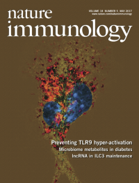
No. 5 May 2017
The nucleic acid sensor TLR9 needs to be tightly regulated to prevent activation by self-ligands in LAMP1+ endolysosomes. Saveanu and colleagues (p 509) identify the aminopeptidase IRAP as a regulator of endosomal trafficking that reduces spurious TLR9 activation. The original image by Loredana Saveanu depicts confocal microscopy imaging of TLR9–GFP (green) with LAMP1+ endosomes (red) in bone-marrow-derived dendritic cells (blue nuclei stained with DAPI). Artwork by Lewis Long.
-
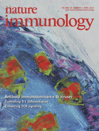
No. 4 April 2017
Although the hierarchical T cell responses induced by infection or immunization have been studied extensively, little is known about the hierarchies of B cell immunodominance. Yewdell and colleagues (p 456; News and Views by Jacob, p 367) define the hierarchy of B cell immunodominance to the five main antigenic sites of the globular domain of hemagglutinin from influenza A virus. The original confocal image by Heather Hickman shows germinal centers (green) in the mediastinal lymph nodes 14 days after respiratory infection with influenza virus. Artwork by Lewis Long.
-
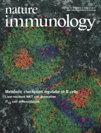
No. 3 March 2017
The metabolic control of B cell fate is unclear. Rickert and colleagues (p 303) show that the kinase GSK3 is a metabolic checkpoint regulator in B cells. The original image by Julia Jellusova shows a mouse spleen 7 days after immunization with sheep red blood cells. Artwork by Lewis Long.
-
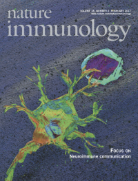
No. 2 February 2017
The nervous and immune systems communicate and reciprocally influence their functional responses. This month's joint focus presents review articles that examine how the nervous system and immune cells interact during development, homeostasis and in pathogenic disease states. http://www.nature.com/focus/neuroimmune_communication/index.html Artwork by Lewis Long.
-
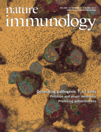
No. 1 January 2017
The cellular sources of the cytokine IL-6 that induces the differentiation of the TH17 subset of helper T cells are unclear. Korn and colleagues (p 74; News and Views by Quintana, p 8) now show that Sirpα+ dendritic cells trans-present IL-6 to T cells through a process that requires the IL-6 receptor subunit IL-6Rα bound to dendritic cells. The original immunofluorescence image by Marina Herwerth shows staining of IL-6 (green) in co-cultures of T cells (white nuclei; DNA-binding dye DAPI) and bone-marrow-derived dendritic cells (red cytoplasm; DAPI). Artwork by Lewis Long.