
Thank you for visiting nature.com. You are using a browser version with limited support for CSS. To obtain the best experience, we recommend you use a more up to date browser (or turn off compatibility mode in Internet Explorer). In the meantime, to ensure continued support, we are displaying the site without styles and JavaScript.

Volume 7 Issue 11, November 2004

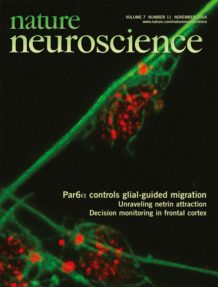
During development, neurons migrate along glial fibers to take their place within the cortical layers. Hatten and colleagues show that overexpression of mPar6α disrupts the coordinated cytoskeletal mechanisms that enable this migration. The cover image shows young cerebellar granule cells migrating along Bergmann glia fibers. The centrosome and nuclei are labeled with dynein intermediate chain (red), and the perinuclear microtubule cage is marked with beta-tubulin (green). In the migrating neurons, the centrosome is positioned forward of the nucleus. Image rendered by Nick Didkovsky. pp 1169 and 1195

Editorial

Top of page ⤴

Book Review

Top of page ⤴

News & Views

  • The perception of a stimulus can result in an emotional response, as we all know, but modulation of perception by emotion has been more difficult to demonstrate. A new study combines imaging and patient data to point to an anatomical substrate for such an effect, raising important implications regarding how sensory-processing impairments might arise in affective disorders.

    • Ralph Adolphs
    News & Views
  • Newborn neurons in the cerebellum migrate along radial glial processes through a series of distinct steps. A report in this issue uses live imaging to grant us a close-up view of the cytoskeletal structures and regulating proteins involved in this migration.

    • Benjamin Adam Samuels
    • Li-Huei Tsai
    News & Views
  • In humans, recollection and familiarity represent qualitatively distinct kinds of memory. A recent study in Nature applied methods commonly used in human research to rats and suggests that their recognition memory may consist of similarly distinct components.

    • Richard G M Morris
    • Michael D Rugg
    News & Views
  • The rostral cingulate zone and the orbitofrontal cortex are active when people monitor the consequences of adaptively changing behavior. A new fMRI study distinguishes their functions, implicating them in situations with different contexts and timing.

    • Markus Ullsperger
    • D Yves von Cramon
    News & Views
Top of page ⤴

Perspective

Top of page ⤴

Brief Communication

Top of page ⤴

Article

Top of page ⤴

Erratum

Top of page ⤴

Search

Quick links