Thank you for visiting nature.com. You are using a browser version with limited support for CSS. To obtain
the best experience, we recommend you use a more up to date browser (or turn off compatibility mode in
Internet Explorer). In the meantime, to ensure continued support, we are displaying the site without styles
and JavaScript.
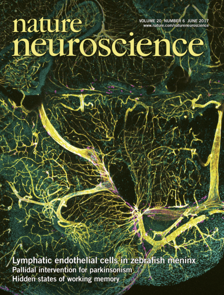
In this issue, Bower and colleagues describe a population of mural lymphatic endothelial cells found along meningeal blood vessels in the adult zebrafish. These mural cells are distinct from meningeal lymphatic vessel cells but form by developmental lymphangiogenesis. They take up low-density lipoproteins from the bloodstream and can modulate angiogenesis during meningeal vascularization. Image: major arteries (yellow), surrounded by mural lymphatic endothelial cells (magenta), enter the zebrafish brain ventrally (nuclei are in cyan), while networks of finer blood capillaries permeate the deeper tissue in this cross-section composite image.p 774
Reproducibility initiatives seek to promote greater transparency and sharing of scientific reagents, procedures and data. Less recognized is the need to share data analysis routines. Nature Neuroscience is launching a pilot project to evaluate the efficacy of sharing code.
Microglia are highly heterogeneous and plastic. However, the dynamics of their turnover have been difficult to visualize. A new multicolor reporter system reveals a plastic but stable network of microglia during health and disease.
Manipulating the activity of a subpopulation of external globus pallidus neurons with optogenetic deep brain stimulation ameliorates motor deficits in a mouse model of Parkinson's disease.
Two groups demonstrate the importance of inputs from the amygdala to the medial prefrontal cortex for signaling aversion across a range of behaviors and motivational drives.
Keeping a picture in mind requires many brain cells to actively communicate ... or does it? There might be more to working memory than neuronal chatter, and silent processes could be hiding right beneath the surface.
Computational techniques are central in many areas of neuroscience and are relatively easy to share. This paper describes why computer programs underlying scientific publications should be shared and lists simple steps for sharing. Together with ongoing efforts in data sharing, this should aid reproducibility of research.
Bower et al. describe a population of mural lymphatic endothelial cells found along meningeal blood vessels in the adult zebrafish. These mural cells are distinct from meningeal lymphatic vessel cells but form by developmental lymphangiogenesis. They take up low-density lipoproteins from the bloodstream and can modulate angiogenesis during meningeal vascularization.
The authors report that genetic disruption of the connectivity of CCK+ basket cells during development reveals a critical role for these interneurons in the regulation of theta oscillatory activity and in the coding of spatial information in the adult mouse hippocampus.
Microglia can expand and divide quickly in the context of CNS pathology, but little is known about the kinetics and clonality of microgliosis. Prinz and colleagues develop a new fate mapping system to monitor microglial dynamics. Microglial self-renewal is found to be a stochastic process under steady state conditions, whereas clonal expansion is observed during disease.
Touch-evoked dynamic mechanical pain is one of most bothersome and prevalent symptoms in chronic pain patients. Here the authors have genetically identified a population of spinal excitatory neurons that contribute to this form of pain. These cells process information from low-threshold Aβ mechanoreceptors and are part of a morphine-resistant pathway.
The external globus pallidus (GPe) is a key contributor to motor suppressing pathways in the basal ganglia. The authors show that optogenetic interventions targeted to specific neuronal subpopulations in the GPe can disrupt pathological activity in the basal ganglia and restore movement for hours beyond stimulation.
Little is known about the mechanisms underlying the orchestration of competing motivational drives. During the simultaneous presentation of cues associated with shock or sucrose, when rats may engage in fear- or reward-related behaviors, amygdala neurons projecting to prefrontal cortex more accurately predict behavioral output and bias animals toward fear-related behavior.
Fear-related disorders are thought to reflect strong and persistent fear associations. The authors show that optogenetic high-frequency stimulation of direct amygdala inputs to the prefrontal cortex can destabilize fear memories and facilitate the extinction of previously acquired fear associations.
The authors optogenetically suppressed CA1 pyramidal neurons during awake sharp-wave ripples (SPW-R) as mice were learning reward locations in a multiwell maze. Comparison of place cells’ activities before and after SPW-R manipulation suggests that interference with SPW-R-associated activity during learning prevents stabilization and refinement of the hippocampal map.
Using calcium imaging and optogenetic manipulation in mice performing a working memory task, the authors show that delay activity in prefrontal cortex pyramidal neurons is crucial for task performance. Optogenetic activation of VIP interneurons enhances the neuronal representation of task-relevant information and improves the animal's memory retention.
Wolff and colleagues show that ‘activity-silent’ brain states are important to working memory. Using a perturbation method to ‘ping’ the brain, they uncover hidden neural states that reflect temporary information held in mind and predict memory performance. They argue that dynamic hidden states could underpin working memory.
The authors show that during sleep, dreaming and specific perceptual dream contents can be localized to a posterior hot zone of the brain. By monitoring activity in this zone, they were able to predict dreaming in real time with high accuracy.
Crockett et al. used model-based fMRI to investigate the neural basis of decisions to profit from harming others vs. themselves. Most people preferred to harm themselves over others for profit. This moral preference was associated with diminished neural responses in value-sensitive brain regions to profit gained from harming others.
By sectioning and sequencing the prefrontal cortex of humans, chimpanzees and macaques, He et al. compiled comprehensive transcriptome atlases of cortical layers. The study provides scores of previously uncharacterized layer-marker genes and more than a hundred human-specific genes, implying that the human neocortex has evolved more than was previously appreciated.