


Advertisement
-
-

Pouchitis: pathophysiology and management
Pouchitis is a common condition that can occur after intestinal surgery. In this Review, Shen discusses our current understanding of the multifactorial pathophysiology, diagnosis and management of pouchitis, primarily in patients with underlying ulcerative colitis.
-
-


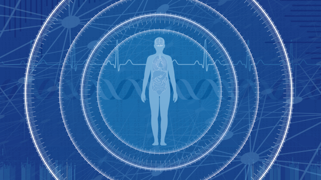
AI in health care
The use of artificial intelligence (AI), including machine learning and large language models, is revolutionizing health care, from drug discovery and development, through to applications in the risk stratification, diagnosis, imaging, monitoring, prognostication, and pharmacological and surgical treatment of patients.
Advertisement
