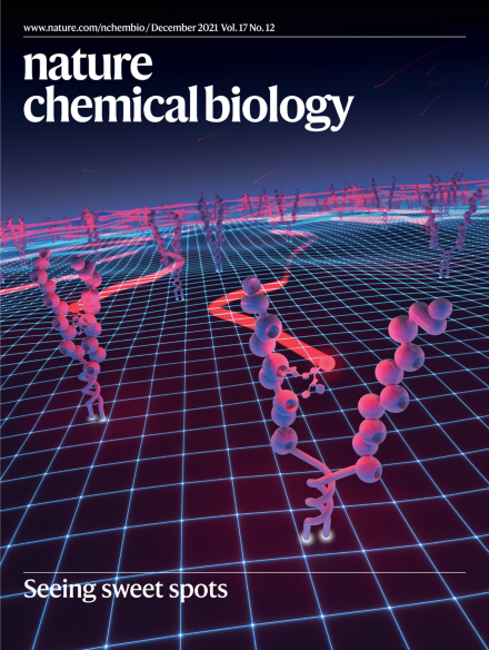
Thank you for visiting nature.com. You are using a browser version with limited support for CSS. To obtain
the best experience, we recommend you use a more up to date browser (or turn off compatibility mode in
Internet Explorer). In the meantime, to ensure continued support, we are displaying the site without styles
and JavaScript.
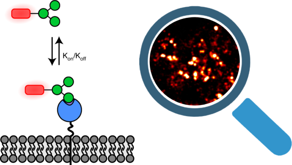
Conceptual visualization of the Glyco-PAINT method. Sugar molecules tagged with a fluorescent marker (in red) bind reversibly to the mannose receptors anchored on the cell membrane, which allows tracking of their movements and provides information on binding affinity and receptor mobility in living cells.
Targeting cholecystokinin receptors (CCKRs) signaling has become an attractive therapeutic strategy for many diseases. The description of cryo-EM structures of CCKRs in the active or inactive states reveal the molecular mechanism of ligand recognition and G-protein-coupling promiscuity.
Chemoproteomics-enabled identification of the targets of a hydroxylated cholesterol metabolite, 20(S)-hydroxycholesterol, reveals it to be the first reported endogenous ligand of the sigma-2 receptor, a drug target for neurodegeneration and neuropathic pain.
Prokaryotic genomes are rife with highly similar toxin–antitoxin modules that need to be protected and diversified at the same time. Studies of paralogous modules now reveal how evolution secures insulation between related systems, without compromising toxin inhibition.
Controlled positioning of the mitotic spindle is key to tissue development and homeostasis. A recent study uncovered an EB1 crotonylation event that orchestrates microtubule dynamics in late mitosis, thus ensuring correct division orientation in vertebrate cells.
This Review surveys how resistance mutations to small molecules and recent mutagenesis technologies have driven biological discovery across diverse fields of inquiry.
Structures of human cholecystokinin receptors in complex with various ligands or G-proteins reveal how different ligand types are recognized and the basis of peptide selectivity in this receptor family, and suggest a stepwise activation mechanism.
Cryo-EM structures of sulfated cholecystokinin 8 bound to the cholecystokinin A receptor in complex with Gs, Gi and Gq heterotrimers reveal structural determinants for G-protein coupling selectivity.
Post-translational site-selective formation of boronoalanine in proteins enables applications of boron for binding partner capture, footprinting of interactions with reactive oxygen species, proteolytic control and mapping of transient structures.
The development of split-engineered base editors (seBEs) enables small-molecule control over DNA deaminase activity, decreasing off-target effects and offering a generalizable solution for temporal control over precise genome editing.
A chemoproteomics profile of the human metabolite 20(S)-hydroxycholesterol exposes its broad connections to the immune system and cancer, revealing it to be a highly selective ligand for the orphan receptor Tmem97 (the σ2 receptor).
Glyco-PAINT, a super-resolution microscopy approach that images fluorescently tagged glycan interactions with lectins, enables the measurement of sugar–lectin complex densities, residence times and diffusion on cell surfaces.
Structural analysis of the Pepper aptamer in complex with its cognate HBC or HBC-like color variants reveals that it binds fluorophore molecules via one non-G-quadruplex base quadruple and one noncanonical G·U base pair.
Structural characterization of three paralogous toxin–antitoxin complexes illuminates how each antitoxin specifically targets its cognate toxin, including auxiliary neutralization interfaces that confer evolvability while preventing loss of activity.
During the biosynthesis of triacsin, the two N–N bond formation reactions necessary to create the unique N-hydroxytriazene moiety are catalyzed by a glycine-utilizing hydrazine-forming enzyme and a nitrite-utilizing N-nitrosating enzyme.
TIP60-mediated crotonylation of the microtubule plus-end tracking protein EB1 at Lys66 regulates attachment of astral microtubules to the lateral cell cortex and spindle positioning.