Thank you for visiting nature.com. You are using a browser version with limited support for CSS. To obtain
the best experience, we recommend you use a more up to date browser (or turn off compatibility mode in
Internet Explorer). In the meantime, to ensure continued support, we are displaying the site without styles
and JavaScript.
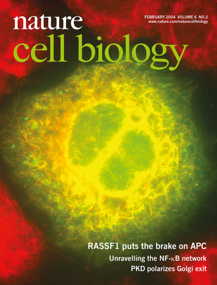
The tumour suppressor RASSF1A localizes to microtubules. Cells expressing HA-tagged RASSF1A (green), were co-stained with anti-b-tubulin (red). cover design: James McQuat
In the post-genome era, there is increasing interest in developing quantitative models of cell function at the molecular level. Although data describing the role of individual proteins in cell behaviour remain rate-limiting for such efforts, large-scale screening approaches are providing results that can be used to map the protein–protein interactions of a living biological system.
Genotoxic stress induces a survival response by releasing the transcription factor NF-κB from its inhibitor IκBα. In response to DNA damage, this initially requires the nuclear modification of the IκB kinase regulator NEMO/IKKγ by SUMO and ubiquitin.
Specific gene expression patterns must be tightly regulated for eukaryotic cells to acquire unique structural and functional features. A key step is transcriptional initiation, during which epigenetic activators are thought to facilitate the process by establishing and maintaining transcription-competent chromatin structures. Accumulating evidence shows that an epigenetic regulator can also control the next step — transcriptional elongation.
The kinetochore attaches the centromeric region of chromosomes to spindle microtubules, allowing equatorial chromosome segregation during mitosis. Proteomic analyses suggest that the kinetochore is a supramolecular complex containing more than 65 proteins that surpasses the size of a ribosome. From within this complex web, stable protein sub-complexes have been extracted, revealing the building blocks for kinetochore assembly.