
Volume 14 Issue 1, January 2021
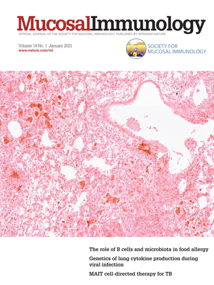
This micrograph shows iron accumulation in the lung of a Mycobacterium tuberculosis - infected C57BL/6 mouse. Iron staining (dark brown) is seen in areas of inflammatory cell infiltration and was markedly decreased in the lungs of mice treated with a pharmacological inhibitor of heme oxygenase-1 compared to those of non-treated animals. The lower intracellular iron accumulation in inhibitor treated mice was associated with increased NOS2 expression and NO production by infected macrophages in response to IFNγ activation as well enhanced bacterial control. For further information, please see the article published in this issue, pp. 253.
Comment
-
Advertisement
