
Figure 15 - Method of endoscopic cricopharyngeal myotomy.
From the following article
Surgical intervention and treatment of oral, pharyngeal motor disorders
Eugene A. Chu and James H. Kelly
GI Motility online (2006)
doi:10.1038/gimo51
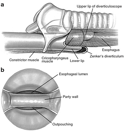
a: Diverticuloscope advanced so upper lip is within esophagus and lower lip is within diverticulum. b:View through diverticuloscope. Cautery, laser, or a stapling device is used to divide the common party wall between the outpouching and the esophagus. (Source: Shapiro J, Van Overbeek J. Endoscopic laser cricopharyngeal myotomy. Oper Tech Otolaryngol–Head Neck Surg 1997;8(4):209-212 with permission.)
Powerpoint slides for teaching
If the slide opens in your browser, Select "File > Save as" to save it.
Download Power Point slide (1,046K)