Figures, tables and video
From the following article
Pharyngeal and esophageal diverticula, rings, and webs
Julia J. Liu and Peter J. Kahrilas
GI Motility online (2006)
doi:10.1038/gimo41
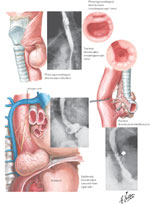
Figure 1
Illustration and radiological appearance of Zenker's mid esophageal and epiphrenic esophageal diverticula.
Full size figure and legend (314K)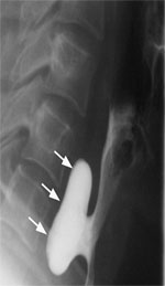
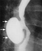
Figure 3
Barium swallow of a patient with midthoracic or traction diverticulum.
Full size figure and legend (31K)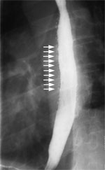
Figure 4
Barium swallow of a patient with intramural pseudodiverticulosis.
Full size figure and legend (28K)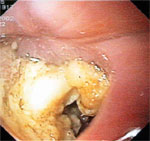
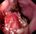
Figure 6
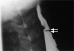
Postendoscopic resection appearance of the Zenker's diverticulum.
Full size figure and legend (58K)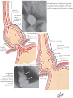
Figure 7
Illustration of A ring (muscular ring) and B ring (Schatzki mucosal ring) in the lower esophagus.
Full size figure and legend (281K)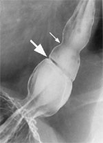
Figure 8
Barium swallow of a patient with A and B rings of the distal esophagus.
Full size figure and legend (40K)