
Volume 222 Issue 1, 13 January 2017
The SEM series: Tongue surface
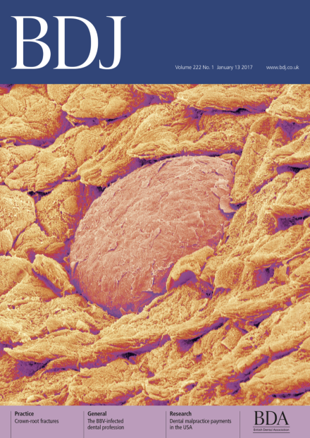
Each cover in volume 222 of the BDJ will feature a scanning electron microscope image showing bacteria or tissues found in the mouth. Scanning electron microscopes (SEMs) allow us to look at surfaces in nano-dimensions, 100 million times smaller than the human eye can see. They work by using beams of electrons instead of light rays. These electrons bounce off the surface of the material in question and are directed at a screen (like a cathode-ray TV screen) where they display a picture of the surface.
This issue features the surface of a tongue. The SEM image shows the filiform papillae covering the majority of the tongue's surface. These are the backward facing scale-like projections and sense pressure on the tongue. The round area (centre) is a fungiform papilla, which contains taste buds.
Credit: SUSUMU NISHINAGA/Science Photo Library
Editorial
-
Advertisement
