Volume 15
-
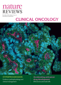
No. 12 December 2018
Immunofluorescence image of a patient-derived colon cancer organoid grown in a 3D in vitro matrix, recapitulating the tumour microanatomy. A strong apical-basal luminal polarity and multiple mitoses are clearly visible in this image.
-
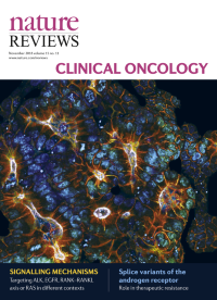
No. 11 November 2018
Immunofluorescence image of a patient-derived colon cancer organoid grown in a 3D in vitro matrix, recapitulating the tumour microanatomy. A strong apical-basal luminal polarity and multiple mitoses are clearly visible in this image.
-
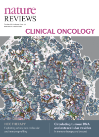
No. 10 October 2018
Immunofluorescence image of a patient-derived colon cancer organoid grown in a 3D in vitro matrix, recapitulating the tumour microanatomy. A strong apical-basal luminal polarity and multiple mitoses are clearly visible in this image.
-
No. 9 September 2018
Immunofluorescence image of a patient-derived colon cancer organoid grown in a 3D in vitro matrix, recapitulating the tumour microanatomy. A strong apical-basal luminal polarity and multiple mitoses are clearly visible in this image.
-
No. 8 August 2018
Immunofluorescence image of a patient-derived colon cancer organoid grown in a 3D in vitro matrix, recapitulating the tumour microanatomy. A strong apical-basal luminal polarity and multiple mitoses are clearly visible in this image.
-
No. 7 July 2018
Immunofluorescence image of a patient-derived colon cancer organoid grown in a 3D in vitro matrix, recapitulating the tumour microanatomy. A strong apical-basal luminal polarity and multiple mitoses are clearly visible in this image.
-
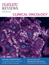
No. 6 June 2018
Immunofluorescence image of a patient-derived colon cancer organoid grown in a 3D in vitro matrix, recapitulating the tumour microanatomy. A strong apical-basal luminal polarity and multiple mitoses are clearly visible in this image.
-
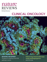
No. 5 May 2018
Immunofluorescence image of a patient-derived colon cancer organoid grown in a 3D in vitro matrix, recapitulating the tumour microanatomy. A strong apical-basal luminal polarity and multiple mitoses are clearly visible in this image. Image supplied by Dr Joseph Regan, Charité— Universitätsmedizin Berlin, Germany
-
No. 4 April 2018
Immunofluorescence image of a patient-derived colon cancer organoid grown in a 3D in vitro matrix, recapitulating the tumour microanatomy. A strong apical-basal luminal polarity and multiple mitoses are clearly visible in this image. Image supplied by Dr Joseph Regan, Charité— Universitätsmedizin Berlin, Germany
-
No. 3 March 2018
Immunofluorescence image of a patient-derived colon cancer organoid grown in a 3D in vitro matrix, recapitulating the tumour microanatomy. A strong apical-basal luminal polarity and multiple mitoses are clearly visible in this image. Image supplied by Dr Joseph Regan, Charité— Universitätsmedizin Berlin, Germany
-
No. 2 February 2018
Immunofluorescence image of a patient-derived colon cancer organoid grown in a 3D in vitro matrix, recapitulating the tumour microanatomy. A strong apical-basal luminal polarity and multiple mitoses are clearly visible in this image. Image supplied by Dr Joseph Regan, Charité— Universitätsmedizin Berlin, Germany
-
No. 1 January 2018
Immunofluorescence image of a patient-derived colon cancer organoid grown in a 3D in vitro matrix, recapitulating the tumour microanatomy. A strong apical-basal luminal polarity and multiple mitoses are clearly visible in this image. Image supplied by Dr Joseph Regan, Charité— Universitätsmedizin Berlin, Germany