Volume 7
-
No. 12 December 2010
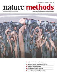
An artistic rendering of a three-dimensional image reconstruction of the superficial mouse brain imaged through a chronic thinned-skull window. In the original image, blood vessels and neurons were visualized using fluorescent markers, and the skull was imaged using second harmonic generation. Here blood vessels are shown in pink, neuronal projections in gray and the skull in white. Original image courtesy of Andy Shih and Phil Tsai; cover by Erin Dewalt. Brief Communication p981
-
No. 11 November 2010
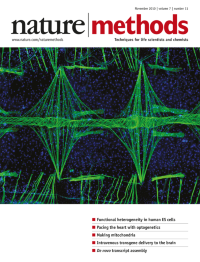
This photograph of an Anopheles gambiae (mosquito) heart took first place in the 2010 Nikon Small world photomicrography competition. The image was taken by Jonas King of the Vanderbilt University Department of Biological Sciences. The heart musculature was stained using Alexa Fluor 488–conjugated phalloidin, and DNA was stained with Hoechst 33342. Other images from this year's competition are on display at http://www.nikonsmallworld.com/.
-
No. 10 October 2010
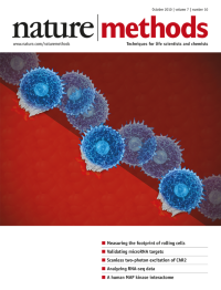
The cover is an artistic depiction of cellular rolling along vascular endothelial cells. Three-dimensional reconstructions of quantitative 'footprint' data from neutrophils were provided by Klaus Ley and Prithu Sundd. Cover design by Erin Dewalt. Brief Communication p821
-
No. 9 September 2010
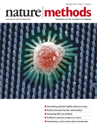
Scanning electron micrograph of a human mesenchymal stem cell grown on an array of posts 12.9 micrometers high. Cover by Erin Dewalt, based on an image provided by Christopher Chen. Brief Communication p733
-
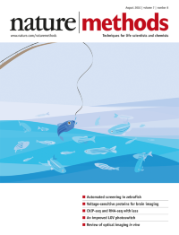
No. 8 August 2010
An artistic interpretation of screening in zebrafish. Cover design by Erin Dewalt, based on an image provided by M. Fatih Yanik and a concept from Craig Millman. Brief Communication p634
-
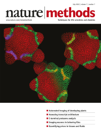
No. 7 July 2010
The cover image shows Arabidopsis thaliana inflorescence meristems expressing fluorescent protein reporters and stained with a vital dye. The image is a blend between raw confocal microscopy data and a three-dimensional rendering of cells segmented using the MARS algorithm. Cover design by Erin Dewalt based on an image provided by Christophe Godin. Article p547, News and Views p506
-
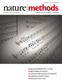
No. 6 June 2010
A DNA sequence put to music. A, C and G are represented by the notes of the same name. For T, the solfege syllable 'ti' is used, which is E in the key of F major (chosen because it includes the other three pitches as well). All of the notes are quarter notes except for the methyl C (represented by a half note with a fermata) and notes in the vicinity of a methyl C, where dotted quarters and eighth notes create syncopation and rhythmic variation. For a trumpet rendition of the piece, please visit Methagora. Cover idea courtesy of Pacific Biosciences; cover design by Erin Dewalt. Article p461
-
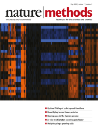
No. 5 May 2010
Depiction of comparative genome hybridization array data for some of the novel insertions identified by Kidd et al. in this issue, as modified by Erin Dewalt. Resource p365
-
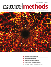
No. 4 April 2010
Modified fly-through rendering of an endoscopy image of blood vessels in the descending colon of a mouse. The blood vessels were labeled by intravenous injection of fluorescent dextran. Original image provided by Seok Yun and modified by Erin Dewalt. Brief Communication p303
-
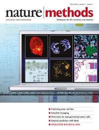
No. 3 March 2010
The cover image shows a range of data visualizations currently used by life scientists. Source images come from figures in the Nature Methods supplement "Visualizing biological data" and from Nature Cell Biology and Nature Biotechnology. Cover design by Seán O'Donoghue and Bang wong. Supplement Foreword p193
-
No. 2 February 2010
Artistic rendering of an electron micrograph of a methlyamine–enriched metagenomic community from a Lake Washington sediment. Sample was obtained by Marina Kalyuzhnaya and Ludmila Chistoserdova; copyright for the original image, Dennis Kunkel Microscopy, Inc., colorization by Ekaterina Latypova. Cover art by Joseph Hiatt. Brief Communication p119
-
No. 1 January 2010
Induced pluripotency is beginning to show its mettle as a powerful tool for biological discovery and is Nature Methods' pick for Method of the Year 2009. Cover design by Erin Dewalt. Special feature starts on p17.
Special